. 2025 Mar 17;16(1):2277.
doi: 10.1038/s41467-025-57456-8.

A CPC-shelterin-BTR axis regulates mitotic telomere deprotection

Affiliations

A CPC-shelterin-BTR axis regulates mitotic telomere deprotection

Diana Romero-Zamora et al. Nat Commun. .

Abstract

Telomeres prevent ATM activation by sequestering chromosome termini within telomere loops (t-loops). Mitotic arrest promotes telomere linearity and a localized ATM-dependent telomere DNA damage response (DDR) through an unknown mechanism. Using unbiased interactomics, biochemical screening, molecular biology, and super-resolution imaging, we found that mitotic arrest-dependent (MAD) telomere deprotection requires the combined activities of the Chromosome passenger complex (CPC) on shelterin, and the BLM-TOP3A-RMI1/2 (BTR) complex on t-loops. During mitotic arrest, the CPC component Aurora Kinase B (AURKB) phosphorylated both the TRF1 hinge and TRF2 basic domains. Phosphorylation of the TRF1 hinge domain enhances CPC and TRF1 interaction through the CPC Survivin subunit. Meanwhile, phosphorylation of the TRF2 basic domain promotes telomere linearity, activates a telomere DDR dependent on BTR-mediated double Holliday junction dissolution, and leads to mitotic death. We identify that the TRF2 basic domain functions in mitosis-specific telomere protection and reveal a regulatory role for TRF1 in controlling a physiological ATM-dependent telomere DDR. The data demonstrate that MAD telomere deprotection is a sophisticated active mechanism that exposes telomere ends to signal mitotic stress.


Conflict of interest statement

Competing interests: The authors declare no competing interests.

Figures

Fig. 1
Fig. 1. TRF1 interactomics reveal CPC and BLM function in MAD telomere deprotection.
a Immunoblots of whole cell extracts from Flag-APEX2 or Flag-APEX2-TRF1 expressing HeLa (representative of n = 3 biological replicates). b Combined telomere fluorescence in situ hybridization (FISH, TelC) and streptavidin (SA) labelling in Flag-APEX2 or Flag-APEX2-TRF1 expressing HeLa, ± APEX2 activation by biotin-phenol (representative of n = 3 biological replicates). Scale bar, 10 µm. c Interactomics timeline. d Volcano plots from the Flag-APEX2-TRF1 interactomics described in (c) and Supplementary Fig. 1c, d. Proteins enriched from Flag-APEX2-TRF1 samples were compared to Flag-APEX2 at respective timepoints and plotted as Log2 fold-change and –Log10 p-value. Enriched CPC (red) and Shelterin (blue) components are indicated (one-sided student’s T-test, n = 5 biological replicates). e Immunoblots of endogenous TRF1 immuno-precipitates from HT1080 6TG. Where indicated, cells were treated for 16 h with 150 ng mL−1 Nocodazole (Noc) immediately prior to sample collection (representative of n = 3 biological replicates). f Hierarchical clustering of proteins with DNA helicase activity (GO:0003678) detected by Flag-APEX2-TRF1 interactomics. Colours indicate Log2-fold change comparing APEX2-TRF1 and APEX2 samples. Helicases with mitotic enrichment are indicated by magenta and RECQL helicases with a blue box. g Immunoblots of whole cell extracts from IMR90 E6E7 hTERT expressing Control or BLM shRNAs 5 days post shRNA transduction (representative of n = 3 biological replicates). h Metaphase-telomere deprotection induced foci (TIF) assays using combined γH2AX immunofluorescence (red) and telomere FISH (TelC, green) in Control or BLM shRNA IMR90 E6E7 hTERT. Cells were treated with 100 ng mL−1 colcemid for the indicated time before sample collection (representative from n = 3 biological replicates). Scale bar, 10 µm, the DNA is stained with DAPI (blue). i Non-telomeric (nonTEL) DNA damage foci and metaphase-TIF (TEL) in shRNA transduced IMR90 E6E7 hTERT treated with 2 or 24 h of 100 ng mL−1 colcemid (all data points from n = 3 biological replicates of 15 or 30 metaphases per replicate for 2 h and 24 h colcemid, respectively, combined in a Tukey box plot, unpaired two-tailed Mann-Whitney U test). Source data are provided as a Source Data file.
Fig. 2
Fig. 2. MAD telomere deprotection requires TRF1 phosphorylation by AURKB.
a TRF1 domain structure and the mutant alleles used in this study. Predicted AURKB sites in the hinge region and BLM binding domain are shown. The deleted amino acids within the FLAG-TRF1ΔBLM variant are indicated in red. b, c Metaphase-telomere deprotection induced foci (TIF) following 24 h of 100 ng mL−1 colcemid in IMR90 E6E7 hTERT expressing Control or TRF1 shRNA and vector or shRNA-resistant TRF1 alleles (all data points from n = 3 biological replicates of 30 metaphases per replicate combined into a Tukey boxplot, Kruskal-Wallis followed by Dunn’s multiple comparisons test). d Below, representative immunoblots of Flag immuno-precipitates from TRF1 shRNA HT1080 6TG cells expressing shRNA-resistant WT Flag-TRF1. Cells were synchronized with a thymidine block, released, and treated with 150 ng mL−1 nocodazole (Noc) where indicated, in the presence or absence of indicated kinase inhibitors for 16 h. The pTRF1-T358 band is indicated with an arrow. Above, quantitation of normalized anti-TRF1-pT358 immunoblot signal (mean +/- s.e.m., n = 3 biological replicates). e Above, example of an anti-TRF1-pT358 immunoblot measuring in vitro AURKB kinase assay on purified Flag-TRF1. Below, quantitation of anti-TRF1-pT358 intensity and the resulting Km (mean +/- s.e.m., n = 3 biological replicates, Km is 0.39 (95% CI: 0.2–0.74) fmol). Source data are provided as a Source Data file.
Fig. 3
Fig. 3. Survivin binds to TRF1 phosphorylated on Ser354 and Thr358.
a Schematic of TRF1 peptide pull-down and subsequent LC-MS/MS analysis for the experiment in (b, c) and Supplementary Fig. 3a. To enrich for mitotic arrest, HT1080 6TG cells were synchronised with a thymidine block and released in the presence of 150 ng mL−1 Nocodazole for 16 h before sample collection and extract preparation. TRF1 peptides immobilized onto beads were incubated with HT1080 6TG mitotic extracts before LC-MS/MS sample preparation and analysis. Drawing of the mass spectrometer was created in BioRender (Cesare, T. (2025) https://BioRender.com/r86h834). b Summary of peptides used for pull-down analysis in (c) and Supplementary Fig. 3a. Red indicates S296, S354, T358, and S367. c Log2-fold change in Chromosome Passenger Complex proteins recovered from mitotically arrested HT1080 6TG extracts by TRF1 phosphopeptides (mean +/- s.e.m., n = 3 biological replicates). d Immunoblots of anti-Flag immuno-precipitates from TRF1 shRNA HT1080 6TG expressing the indicated shRNA-resistant Flag-TRF1-WT or mutant alleles. Cells were synchronised with a thymidine block, released, and treated with 150 ng mL−1 of nocodazole for 16 h. TRF1-DHD is a deletion of residues 336–367 (representative of n = 3 biological replicates is shown). Source data are provided as a Source Data file.
Fig. 4
Fig. 4. AURKB phosphorylates the TRF2 basic domain to promote MAD telomere deprotection.
a TRF2 domain structure and the mutant alleles used in this study. Predicted AURKB sites in the basic domain are indicated. b Below, representative immunoblots of Myc immuno-precipitates from TRF2 shRNA HT1080 6TG expressing Myc-TRF2-WT. Cells were synchronized with a thymidine block, released in the presence or absence of 150 ng mL−1 nocodazole (Noc) and the indicated kinase inhibitors for 16 h. Above, quantitation of normalized anti-TRF2-pS65 immunoblot signal (mean +/- s.e.m., n = 3 biological replicates). c Left, example of anti-TRF2-pS65 immunoblot measuring in vitro AURKB kinase assay on purified Myc-TRF2. Right, quantitation of anti-TRF2-pS65 intensity and the resulting Km (mean +/- s.e.m., n = 3 biological replicates, Km is 1.3 (95% CI: 0.49–4.0) fmol). d, e Quantitation of interphase-telomere deprotection induced foci (TIF) (d) and metaphase-TIF (e) in TRF2 shRNA IMR90 E6E7 hTERT expressing TRF2-WT or the indicated TRF2 alleles. For (d) Above, all data points from n = 3 biological replicates of 45 nuclei per replicate compiled into a Tukey boxplot, Kruskal-Wallis followed by Dunn’s multiple comparisons test. Below, representative immunoblots of whole-cell extracts derived from the cell cultures used in this experiment. For (e), cells were treated with 2 or 24 h of 100 ng mL−1 colcemid (all data points from n = 3 biological replicates of 15 and 30 metaphases per replicate for 2 h and 24 h colcemid, respectively, compiled into a Tukey boxplot, Kruskal-Wallis followed by Dunn’s multiple comparisons test). Source data are provided as a Source Data file.
Fig. 5
Fig. 5. TRF2 basic domain modification during mitotic arrest promotes t-loop opening.
a, b Interphase-telomere deprotection induced foci (TIF) (a) and Metaphase-TIF (b) in Trf2F/F Cre-ER LgT MEFs expressing TRF2-WT or the indicated TRF2 alleles. Where indicated, endogenous mTrf2 was deleted by 4-Hydroxytamoxifen (4-OHT) addition 36 h prior to sample fixation. In (b) cells were treated with 14 h of 400 ng mL−1 Nocodazole prior to sample preparation (all data points from n = 3 biological replicates of ≥ 50 cells (a) or 30 metaphases (b) per replicate, compiled into a Tukey box plot, Kruskal-Wallis followed by Dunn’s multiple comparisons test). c Representative examples of telomere macromolecular structure as visualized by AiryScan microscopy from Trf2F/F Cre-ER LgT MEFs treated with 400 ng mL−1 Nocodazole for 14 h and collected by mitotic shake-off. Samples were trioxsalen cross-linked in situ, and the chromatin spread on coverslips through cytocentrifugation before telomere FISH labelling. Scale bar, 2 µm. Representative of n = 3 biological replicates. d Quantification of looped telomeres from Trf2F/F Cre-ER LgT MEFs expressing the indicated TRF2 alleles in mitotically arrested samples prepared and shown as in (c) and Supplementary Fig. 5c. Where indicated, endogenous mTrf2 was deleted by 4-OHT addition 36 hours prior to sample fixation (mean +/- s.e.m., n = 3 biological replicates of 200 telomeres per replicate, Ordinary one-way ANOVA followed by Šídák’s multiple comparisons test, F = 6.463, DF = (6, 14)). Source data are provided as a Source Data file.
Fig. 6
Fig. 6. TRF2 phosphorylation promotes BTR-dependent MAD telomere deprotection.
a BLM domain structure and the mutant alleles used in this study. The compromised function of the mutant alleles is indicated. b, c Above, Metaphase-telomere deprotection induced foci (TIF) following 24 h of 100 ng mL−1 colcemid in IMR90 E6E7 hTERT expressing Control or BLM shRNA and shRNA-resistant BLM alleles (all data points from n = 3 biological replicates of 30 metaphases per replicate, compiled into a Tukey Box Plot, Kruskal-Wallis followed by Dunn’s multiple comparisons test). Below: representative immunoblots of whole cell extracts derived from the untreated cell cultures used in this experiment. d TOP3A domain structure and the mutant allele used in this study. e Above, Metaphase-TIF following 24 h of 100 ng mL−1 colcemid in IMR90 E6E7 hTERT fibroblasts expressing Control or TOP3A shRNA and vector or shRNA-resistant TOP3A alleles (all data points from n = 3 biological replicates of 30 metaphases per replicate compiled into a Tukey Box Plot, Kruskal-Wallis followed by Dunn’s multiple comparisons test). Below, representative immunoblots of whole cell extracts derived from the untreated cell cultures used in this experiment. f Above, Metaphase-TIF following 24 h of 100 ng mL−1 colcemid in IMR90 E6E7 hTERT expressing Control, BLM or TRF2 shRNA, and vector or TRF2-2D (S62D and S65D) (all data points from n = 3 biological replicates of 30 metaphases per replicate compiled into a Tukey Box Plot, Kruskal-Wallis followed by Dunn’s multiple comparisons test). Below, representative immunoblots of whole cell extracts derived from the untreated cell cultures used in this experiment. Source data are provided as a Source Data file.
Fig. 7
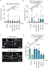
Fig. 7. TRF2 phosphorylation promotes mitotic death during mitotic arrest.
a Mitotic duration for TRF2 shRNA IMR90 E6E7 hTERT expressing the indicated TRF2 alleles. Cells were observed for 50 h in cultures treated with 100 ng mL−1 colcemid (median of N cells from n = 2 experimental replicates). b Representative captures from phase contrast live imaging in (a). Mitotic outcomes are indicated. Time is shown as hours: minutes relative to mitotic entry. Scale bar, 20 µm. Representative of n = 2 experimental replicates. c Ratio of mitotic outcomes as shown in (b) after indicated duration of mitotic arrest (N cells from n = 2 experimental replicates). d Number of mitotic cell death and mitotic slippage from (c) in the indicated conditions within 2–6 h of mitotic arrest (n = 2 experimental replicates, unpaired two-tailed Fisher’s Exact test). e Model of MAD telomere deprotection. (1) AURKB phosphorylates TRF1 at S354 and T358; (2) Survivin interacts with pS354 and pT358 to promote (3) AURKB phosphorylation of TRF2 at S62 and S65, enabling (4) BTR to dissolve t-loops. Source data are provided as a Source Data file.

References

    1. de Lange, T. Shelterin-mediated telomere protection. Annu. Rev. Genet.52, 223–247 (2018). - PubMed
    1. Cesare, A. J. & Karlseder, J. A three-state model of telomere control over human proliferative boundaries. Curr. Opin. Cell Biol.24, 731–738 (2012). - PMC - PubMed
    1. Karlseder, J., Smogorzewska, A. & de Lange, T. Senescence induced by altered telomere state, not telomere loss. Science295, 2446–2449 (2002). - PubMed
    1. d'Adda di Fagagna, F. et al. A DNA damage checkpoint response in telomere-initiated senescence. Nature426, 194–198 (2003). - PubMed
    1. Kaul, Z., Cesare, A. J., Huschtscha, L. I., Neumann, A. A. & Reddel, R. R. Five dysfunctional telomeres predict onset of senescence in human cells. EMBO Rep.13, 52–59 (2011). - PMC - PubMed

MeSH terms