. 2025 Mar 18;11(1):23.
doi: 10.1038/s41421-024-00763-z.

Trans-Golgi network tethering factors regulate TBK1 trafficking and promote the STING-IFN-I pathway

Affiliations

Trans-Golgi network tethering factors regulate TBK1 trafficking and promote the STING-IFN-I pathway

Jinrui Wang et al. Cell Discov. .

Abstract

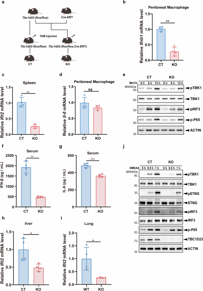
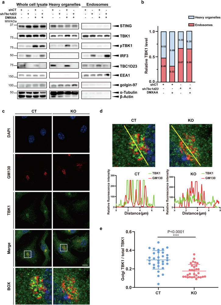
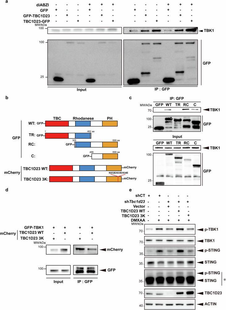
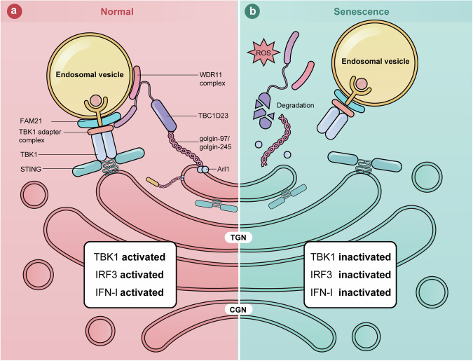
The cGAS-STING pathway mediates the innate immune response to cytosolic DNA, contributing to surveillance against microbial invasion or cellular damage. Once activated, STING recruits TBK1 at the trans-Golgi network (TGN), which in turn phosphorylates IRF3 to induce type I interferon (IFN-I) expression. In contrast to STING, little is known about how TBK1 is transported to the TGN for activation. Here, we show that multiple TGN tethering factors, a group of proteins involved in vesicle capturing, are indispensable for STING-IFN-I signaling. Deletion of TBC1D23, a recently reported tethering factor, in mice impairs the STING-IFN-I signaling, but with insignificant effect on STING-NF-κB signaling. Mechanistically, TBC1D23 interacts with TBK1 via the WASH complex subunit FAM21 and promotes its endosome-to-TGN translocation. Furthermore, multiple TGN tethering factors were reduced in aged mice and senescent fibroblasts. In summary, our study uncovers that TGN tethering factors are key regulators of the STING-IFN-I signaling and suggests that their reduction in senescence may produce aberrant STING signaling.


Conflict of interest statement

Conflict of interest: The authors declare no competing interests.

Figures

Fig. 1
Fig. 1. Multiple TGN tethering factors are reduced in aged mice or upon oxidative stress.
a, b Lungs (a) or hearts (b) from young and aged mice were extracted and analyzed for expression levels of TGN tethering factors by immunoblotting. c Primary mouse lung fibroblasts were first treated with etoposide (50 μM) for 12 h or pretreated with H2O2 (200 μM) for 2 h and continued to be cultured in the presence or absence of etoposide (50 μM) for 12 h. N-acetylcysteine (NAC, 5 mM) was used to remove ROS. Untreated cells were used as a control, and the protein levels of TGN tethering factors were analyzed by immunoblotting. d Statistical analysis of golgin-245 and TBC1D23 expression levels, 30 cells were counted in each group. The levels were determined by normalizing the image gray values in c. Statistical results of golgin-97 and GCC88 are shown in Supplementary Fig. S1. e Immortalized human cerebral microvascular endothelial HCMEC/D3 cells were pretreated with H2O2 (500 μM). Distribution of Golgi proteins was analyzed by fluorescence confocal microscopy. Scale bar = 10 μm. f Quantifying the ratio of clustered protein fluorescence intensity to total fluorescence intensity within a cell (n = 30). One representative experiment of at least three independent experiments is shown. Data analyzed by two-tailed t-test and shown as mean ± SD (n ≥ 3). ns, not significant, p > 0.05; *p < 0.05; **p < 0.01; ***p < 0.001; ****p < 0.0001.
Fig. 2
Fig. 2. Multiple TGN tethering factors promote activation of the cGAS-STING-IFN-I signaling.
a Immortalized human cerebral microvascular endothelial HCMEC/D3 cells were pretreated with H2O2 (200 μM) for 2 h. Endogenous STING activation was induced by diABZI (5 μM), and the phosphorylation levels of TBK1, STING and IRF3 were analyzed by immunoblotting at different time points. b Immortalized human cerebral microvascular endothelial HCMEC/D3 cells were pretreated with H2O2 (200 μM) for 2 h. The mRNA levels of IFNB1 were analyzed by qPCR after diABZI (5 μM) treatment for various times. c HCMEC/D3 cells were processed as in a, and cells at the 1 h time point were analyzed by confocal fluorescence microscopy. Scale bar = 5 μm. d For images in c, Golgi-associated TBK1 phosphorylation intensity (using Golgi-resident protein GM130 as a reference) was counted and ratioed to the whole-cell signal intensity. e, f THP-1 cells stably expressing an inducible gene knockdown system were treated with dox (1 μM) for 72 h and followed by treatment with MnCl2 (200 μM) for 24 h. Cells were collected, and mRNA levels of IFNB1 (e) and TNF-α (f) were determined by qPCR. g, h THP-1 cells stably expressing an inducible gene knockdown system were treated with dox (1 μM) for 72 h and followed by treatment with diABZI (5 μM) for 1 h. Phosphorylation of TBK1 and IRF3 was analyzed by immunoblotting. i Statistical analysis was performed after normalizing the image gray values of the experimental results in g, h. One representative experiment of at least three independent experiments is shown. Data are analyzed by two-tailed t-test and shown as mean ± SD (n ≥ 3). ns, not significant, p > 0.05; *p < 0.05; **p < 0.01; ***p < 0.001; ****p < 0.0001.
Fig. 3
Fig. 3. Tbc1d23 knockout mice exhibit reduced STING-IFN-I signaling.
a Generation of Tbc1d23 conditional knockout mouse. b CT or Tbc1d23 KO mice were injected intraperitoneally with 40 mg/kg of MnJβ adjuvant (1.4 mg/mL) dissolved in saline. Ten hours later, peritoneal macrophages were collected, and the expression levels of Ifnb1 were analyzed by qPCR. c CT or Tbc1d23 KO Mice were injected intraperitoneally with 40 mg/kg of MnJβ adjuvant (1.4 mg/mL) dissolved in saline. Ten hours later, Spleens were collected, and the expression levels of Ifit2 were analyzed by qPCR. d CT or Tbc1d23 KO Mice were injected intraperitoneally with 40 mg/kg of MnJβ adjuvant (1.4 mg/mL) dissolved in saline. Ten hours later, peritoneal macrophages were collected, and the expression levels of Il-6 were analyzed by qPCR. e BMDM cells obtained from CT or Tbc1d23 KO mice were treated with MnCl2 (200 μM) various times, and then subjected to immunoblotting analysis. fi CT or Tbc1d23 KO Mice were injected intraperitoneally with 25 mg/kg of DMXAA (5 mg/mL) dissolved in 7.5% NaHCO3. Three hours later, the levels of serum IFN-β (f) and IL-6 (g) were determined with an ELISA kit. Organs from each mouse were extracted, and the expression levels of Ifit2 were determined by qPCR (h, i). j BMDM cells obtained from CT or Tbc1d23 KO mice were treated with DMXAA (25 μg/mL) for various time points, and then subjected to immunoblotting analysis. One representative experiment of at least three independent experiments is shown. Data analyzed by two-tailed t-test and shown as mean ± SD (n ≥ 3). ns, not significant, p > 0.05; *p < 0.05; **p < 0.01; ***p < 0.001; ****p < 0.0001.
Fig. 4
Fig. 4. TBC1D23 promotes TBK1 translocation to the Golgi apparatus.
a MEFs stably expressing TBC1D23-targeting shRNA (shTbc1d23), or control shRNA (shCT) were treated with DMXAA (25 μg/mL) for 30 min. Endosomes and heavy organelles (Golgi, endoplasmic reticulum, mitochondria, etc.) were isolated, and subjected to immunoblotting. EEA1 and golgin-97 were used to indicate endosomes and Golgi, respectively. b Quantification based on grey values. Results from three different experiments in (a) were combined, and data were averaged. c, d BMDM cells were treated with DMXAA (25 μg/mL) for 30 min. Golgi-association of TBK1 was analyzed by confocal fluorescence microscopy, using Golgi-resident protein GM130 as a reference. Line scans show the related intensity profiles of TBK1 (green) and GM130 (red). Scale bar = 5 μm. e Quantitative analysis of the ratio of Golgi TBK1 to total TBK1, as shown in (c). One representative experiment of at least three independent experiments is shown. Data are analyzed by two-tailed t-test and shown as mean ± SD (n ≥ 3). ns, not significant, p > 0.05; *p < 0.05; **p < 0.01; ***p < 0.001; ****p < 0.0001.
Fig. 5
Fig. 5. TBC1D23 interacts with TBK1 and regulates the STING-IFN-I signaling.
a THP-1 cells stably expressing GFP, GFP-TBC1D23, or TBC1D23-GFP were treated with or without diABZI and subjected to immunoprecipitation using GFP beads. Bound TBK1 was analyzed by immunoblotting. b Schematic diagram of full-length and various truncations of TBC1D23 used in the experiment. Red: TBC domain; Blue: Rhodanese domain: Yellow: PH domain. WT: wild type; TR: TBC and Rhodanese domain; RC: Rhodanese and C terminal PH domain; C: C terminal PH domain; TBC1D23 3 K: TBC1D23 with a K632E/K633E/K634E triple mutant. c HEK293T cells expressed different truncates were subjected to immunoprecipitation using GFP beads. Bound TBK1 was analyzed by immunoblotting. d HEK293T cells were expressed GFP-TBK1 together with TBC1D23 WT-mCherry or 3K-mCherry and subjected to immunoprecipitation using GFP beads. Bound TBK1 was analyzed by immunoblotting. e MEFs stably expressing TBC1D23-targeting shRNA (shTbc1d23), or control shRNA (shCT) were rescued with an empty vector, TBC1D23 WT or 3 K mutant. Cells were treated with DMXAA (25 μg/mL) for 30 min, and then subjected to immunoblotting. *: Long time exposure. One representative experiment of at least three independent experiments is shown.
Fig. 6
Fig. 6. A model showing how TGN tethering factors regulate the STING-IFN-I signaling.
a In normal cells, TGN tethering factors promote STING-IFN-I signaling by mediating endosome-to-TGN translocation of TBK1. b In senescent cells, stresses, such as ROS, lead to the degradation of TGN tethering factors. The endosome-to-TGN translocation of TBK1 is blocked, and STING-IFN-I signaling is inhibited.

References

    1. Ishikawa, H., Ma, Z. & Barber, G. N. STING regulates intracellular DNA-mediated, type I interferon-dependent innate immunity. Nature461, 788–792 (2009). - PMC - PubMed
    1. Hopfner, K.-P. & Hornung, V. Molecular mechanisms and cellular functions of cGAS–STING signalling. Nat. Rev. Mol. Cell Biol.21, 501–521 (2020). - PubMed
    1. Yum, S., Li, M., Fang, Y. & Chen, Z. J. TBK1 recruitment to STING activates both IRF3 and NF-?B that mediate immune defense against tumors and viral infections. Proc. Natl. Acad. Sci. USA118, e2100225118 (2021). - PMC - PubMed
    1. Liu, S. et al. Phosphorylation of innate immune adaptor proteins MAVS, STING, and TRIF induces IRF3 activation. Science347, aaa2630 (2015). - PubMed
    1. Fang, R. et al. NEMO–IKKβ Are Essential for IRF3 and NF-κB Activation in the cGAS–STING Pathway. J. Immunol.199, 3222–3233 (2017). - PubMed

LinkOut - more resources