. 2025 Mar 17;16(1):2615.
doi: 10.1038/s41467-025-57788-5.

Cortico-striatal circuit mechanisms drive the effects of D1 dopamine agonists on memory capacity in mice through cAMP/PKA signalling

Affiliations

Cortico-striatal circuit mechanisms drive the effects of D1 dopamine agonists on memory capacity in mice through cAMP/PKA signalling

Maria De Risi et al. Nat Commun. .

Abstract

Working memory capacity (WMC), the number of items remembered in a short-time interval, is regulated by fronto-striatal dopamine (DA) and is reduced in schizophrenia. We investigated how excessive and insufficient D1 dopamine receptor stimulation impairs and expands WMC, focusing on the cAMP/PKA pathway in the fronto-striatal circuit. Low doses of the D1 agonist SKF 38393 enhance WMC by activating the striatum (mice remember more objects), while high doses, paradoxically, impair WMC, activating the same pathway in the medial prefrontal cortex (mPFC) but inhibiting it in the striatum. This impairment, arising from mPFC-driven recruitment of inhibitory striatal parvalbumin interneurons, can be prevented by optogenetic inhibition of the mPFC-striatal pathway. Low doses of SKF 38393 also rescue WMC deficits in a schizophrenia mouse model. These results highlight the need for a systems pharmacology approach that considers complex brain interactions and intracellular signalling pathways, rather than isolated drug-receptor interactions, to develop memory-enhancing treatments.


Conflict of interest statement

Competing interests: The authors declare no competing interests.

Figures
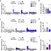
Fig. 1
Fig. 1. SKF 38393 exerts inverted-U dose-response effects on memory capacity.
a Effects of increasing doses of SKF 38393 (0.001, 0.01 and 0.1 mg/kg) on single object exploration (Repeated measures ANOVA: Treatment: F3,40 = 2.68; p = 0.0593; Object F5,200 = 44.28; p < 0.0001; Treatment x Object F15,200 = 3.9; p < 0.0001, Dunnett’s post hoc) and (b) on the discrimination index during the 6 different objects test (6-DOT) (One-way ANOVA: Treatment: F3,40 = 5.42; p = 0.0032, Tukey’s post hoc). c Effects of increasing doses of SKF 38393 (0.001 and 0.01 mg/kg) on single object exploration (Repeated measures ANOVA: Treatment: F2,34 = 2.39; p = 0.1061; Object F7,238 = 8.9; p < 0.0001; Treatment x Object F14,238 = 1.38; p = 0.1628, Dunnett’s post hoc) and (d) on discrimination index during the 8 different objects test (8-DOT) (One-way ANOVA: Treatment: F2,34 = 13.5; p < 0.0001, Tukey’s post hoc). e Effects of SKF 38393 0.1 mg/kg combined with SCH-23390 (0.001 mg/kg) on single object exploration (Repeated measures ANOVA: Treatment: F2,27 = 1.94; p = 0.1631; Object F5,135 = 23.1; p < 0.0001; Treatment x Object F10,135 = 2.3; p = 0.0157, Dunnett’s post hoc) and (f) on the discrimination index during the 6-DOT (One-way ANOVA: Treatment: F2,27 = 5.80; p = 0.0080, Tukey’s post hoc). In the upper panel was reported a scheme of the test phase of the 6 or 8-DOT. The red circle indicates the novel object. The histogram reported the exploration of the novel object (N) versus the familiar ones. Data are mean values  ±  SEM. *p < 0.05 SKF vs vehicle; #p  <  0.05 novel object (N) vs familiar. Source data are provided as a Source Data file.
Fig. 2
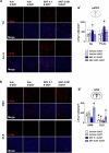
Fig. 2. Improving and impairing doses of SKF 38393 increase c-Fos+ cells in the striatum and the medial prefrontal cortex, respectively.
a, b Representative images of c-Fos-immunofluorescence staining in the (a) cingulate cortex (Cg) and prelimibic/infralimbic cortex (PrL/IL) and (b) the dorso-medial and dorso-lateral striatum (DMS and DLS) of animals tested in the 6-DOT and the 8-DOT injected with vehicle (Veh), the improving dose (0.001 mg/kg), or the impairing dose (0.1 mg/kg) of SKF 38393. a’, b’ Quantification of the number of c-Fos+ cells/mm2 after SKF 38393 treatment in Cg and PrL/IL of medial prefrontal cortex (mPFC) (Repeated measures ANOVA: Treatment: F3,17 = 9.1; p = 0.0008; Subregion: F1,17 = 11.65; p = 0.0033; Treatment x Subregion; F3,17 = 4.5; p = 0.0165, Tukey’s post hoc) and in the DMS and DLS of striatum (STR) (Repeated measures ANOVA: Treatment: F3,17 = 17.9; p < 0.0001; Subregion: F1,17 = 188.4; p < 0.0001; Treatment x Subregion; F3,17 = 10.6; p = 0.0003, Tukey’s post hoc). Brain regions were highlighted with red circles in the upper panel. Data are mean values  ±  SEM. *p < 0.05 SKF vs vehicle; #p < 0.05 DLS vs DMS. Scale bars: 50 µm. Source data are provided in the Source Data file.
Fig. 3
Fig. 3. Improving and impairing doses of SKF 38393 activate PKA/cAMP signalling in the striatum and the medial prefrontal cortex, respectively.
a, b Histograms on the left show the quantification of PKA-phosphorylated substrates, which was expressed as the sum of percentage difference of each band relative to the control. Representative immunoblots of protein extracts from the medial prefrontal cortex (mPFC) and the whole striatum (STR) (red circles in the pictures) probed with a phospho-PKA substrate antibody are reported on the right. Hsc70 was used as a loading control. We observed increased phosphorylation of PKA substrates in the mPFC at the impairing dose (One-way ANOVA: Treatment: F2,20 = 7.69; p < 0.0033, Tukey’s post hoc), while the improving dose increased phosphorylation of PKA substrates in the STR (One-way ANOVA: Treatment: F2,20 = 24.86; p < 0.0001, Tukey’s post hoc). Data are mean values  ±  SEM. *p < 0.05 SKF vs vehicle; #p < 0.05 SKF 0.001 vs SKF 0.1. Source data are provided in the Source Data file.
Fig. 4
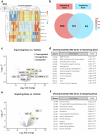
Fig. 4. Phosphoproteomic analysis in the striatum revealed a unique pattern of phosphorylation after a low dose of SKF 38393.
a Heat map of the 50 most variable phosphosites in mice striatum (STR) treated with vehicle (VEH), an improving dose (SKF-LOW), or an impairing dose (SKF-HIGH) of SKF 38393. The phosphosites with increased intensities are marked in red and the phosphosites with lower intensities are marked in blue. b Venn Diagram of differentially expressed phosphosites in mice treated either with vehicle, an improving dose, or an impairing dose. c Volcano plot of the phosphosites belonging to the low improving dose relative to vehicle based on the average ratio of three technical replicates and p-value (− log10 p-value). Grey points represent unchanged proteins, and orange and purple represent the upregulated and downregulated proteins, respectively. The indicated proteins are discussed in the text. d GO terms overrepresented in the low improving versus vehicle comparison. GO:MF refers to Gene ontology Molecular Function, GO:BP refers to the Gene Ontology Biological processes and GO:CC refers to the Gene Ontology Cellular Components. e Volcano plot of the phosphosites belonging to the high impairing dose versus vehicle comparison according to the average ratio of three technical replicates and p-value (− log10 p-value). Grey points represent unchanged proteins, and orange and purple represent the upregulated and downregulated proteins respectively. The indicated proteins are discussed in the text. f GO terms overrepresented in the high impairing dose versus vehicle comparison. GO:MF refers to Gene ontology Molecular Function, GO:BP refers to the Gene Ontology Biological processes and GO:CC refers to the Gene Ontology Cellular Components. Differential abundance of phosphosites analysis was performed using the one way ANOVA test with a threshold of FDR < 0.05. Source data are provided in the Source Data file.
Fig. 5
Fig. 5. Inhibition of striatal D1R downstream signalling prevents the memory capacity increase mediated by SKF 38393, while its stimulation mimics the SKF-improving effects.
a Schematic of the experimental design.Mice were administered systemically with 0.001 mg/kg SKF 38393 together with intra-striatal injection of vehicle or the cAMP antagonist (RP)-cAMPS (Rpc), tested in the 8 different objects test (8-DOT) and subsequently assayed for PKA phosphorylation in the striatum (STR). a’ The discrimination index during the 8-DOT (One-way ANOVA: Treatment: F1,22 = 15.187; p = 0.0008). a” Representative immunoblot and quantification of PKA phosphorylation substrates (One-way ANOVA: Treatment: F1,12 = 20.31; p = 0.0007). b Schematic of the experimental design. Mice were infected with AAV2/5-CaMKIIa-HA-rM4D(Gi)-IRES-mCitrine (AAV-Gi) in the STR, injected withSKF 38393 0.001 mg/kg or CNO,tested in the 8-DOT and subsequently assayed for PKA phosphorylation. b’ Effects of SKF 38393, CNO, or their combination on the discrimination index (One-way ANOVA: Treatment: F2,30 = 10.54; p = 0.0003, Tukey’s post hoc) (b”) Representative immunoblot and quantification of PKA phosphorylation substrates (One-way ANOVA: Treatment: F1,11 = 16.06; p = 0.0021). c Schematic of the experimental design: mice were systemically administered with SKF 38393 0.001 mg/kg or with intra-striatal injection of vehicle or the cAMP agonist (Sp)-cAMPS (Spc). c’ The discrimination index during the 8-DOT under SKF 38393 or Spc treatment (One-way ANOVA: Treatment: F1,22 = 0.02; p = 0.8742) (c”) representative immunoblot and quantification of PKA phosphorylation substrates (One-way ANOVA: Treatment: F1,13 = 0.02; p = 0.8669) were reported. d Schematic of the experimental design. Mice were infected with AAV2/5-CaMKIIa-HA-rM3D(Gs)-IRES-mCitrine (AAV-Gs) in the STR, injected with vehicle or CNO,,tested in the 8-DOT and subsequently assayed for PKA phosphorylation. d’ Effects of vehicle or CNO in animals expressing AAV-Gs on the discrimination index (One-way ANOVA: Treatment: F1,22 = 19.93; p = 0.0002). d” Representative immunoblot and quantification of PKA substrates phosphorylation (One-way ANOVA: Treatment: F1,11 = 8.15; p = 0.0156). Data in bar charts are presented as mean values  ±  SEM. *p < 0.05 vs SKF or vehicle. Scale bars: 100 µm. Source data are provided as a Source Data file.
Fig. 6
Fig. 6. Inhibition of the D1R downstream signalling in the medial prefrontal cortex prevents the memory capacity impairment mediated by the high dose of SKF 38393.
a Schematic of the experimental design. Mice were administered systemically with the impairing dose of SKF 38393 (0.1 mg/kg) or vehicle and injected locally in the medial prefrontal cortex (mPFC) with vehicle or the cAMP antagonist (RP)-cAMPS (Rpc, 0.025 µg/0.3 µl). The mice were then tested in the 6 different objects test (6-DOT) and subsequently assayed for PKA phosphorylation in both mPFC and striatum (STR) (red circles in the pictures). b Effects of the treatment with vehicle or SKF 38393 0.1 mg/kg or intra-mPFC of Rpc or the combination of both. Intra-mPFC Rpc alone did not change the performance in the 6-DOT, compared to vehicle. When given in combination with SKF, it prevented the impairing effects mediated by the high dose of SKF on WMC (One-way ANOVA: Treatment: F3,32 = 4.23; p = 0.0126, Tukey’s post hoc). c, d Representative immunoblots and quantification of PKA substrates phosphorylation. Intra-mPFC Rpc suppresses the PKA substrates phosphorylation increase caused by the impairing dose of SKF 38393in the mPFC (c) (One-way ANOVA: Treatment: F3,23 = 4.95; p = 0.0085, Tukey’s post hoc) and the hypoactivation of PKA in the STR of SKF (d) (One-way ANOVA: Treatment: F3,23 = 6.640; p = 0.0022, Tukey’s post hoc). Data in bar charts are presented as mean values  ±  SEM. *p < 0.05 vs vehicle. #p < 0.05 vs SKF 0.1 mg/kg. Source data are provided in the Source Data file.
Fig. 7
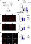
Fig. 7. Optogenetic inhibition of the fronto-striatal circuit rescues the memory capacity impairment induced by the higher dose of SKF 38393 by decreasing Parvalbumin interneuron activation in the striatum.
a Schematic of the experimental design. Mice were infected with AAV2/2-hSyn-eNpHR 3.0-EYFP or AAV2/2-hSyn-EGFP in the medial prefrontal cortex (mPFC) and fiber optics were implanted in the striatum (STR). Photoinhibition was activated after systemic administration of the impairing dose (0.1 mg/kg) of SKF 38393 and during the 6 different objects test (6-DOT). b Discrimination index of vehicle or SKF 38393 0.1 mg/kg injected mice with light OFF and under photoinhibition (light ON). The group treated with SKF 38393 0.1 mg/kg under photoinhibition increased the discrimination index compared to SKF 38393 light OFF and to SKF 38393 light ON without Halorhodopsin (One-way ANOVA: Treatment: F4,54 = 11.13; p < 0.0001; Tukey’s post hoc). cf c-Fos immunostaining and quantification of the number of c-Fos positive cells/mm2 in the mPFC and STR in SKF 38393 0.1 mg/kg light OFF and light ON mice. The optogenetic inhibition did not change the number of c-Fos+ cells in the mPFC (d) but increased it in the striatum (One-way ANOVA: Light: F1,12 = 6.34; p = 0.0270) (f). Scale bars: 100 µm. gh Photoinhibition decreased the number of double-labelled Parvalbumin (PV+) and c-Fos+ neurons (One-way ANOVA: Light: F1,12 = 7.0; p = 0.0209). Arrows indicate double labelled PV+/c-Fos+ neurons. Scale bars: 25 µm. Data in bar charts are presented as mean values  ±  SEM. *p < 0.05 vs SKF #p < 0.05 vs Light OFF. Source data are provided in the Source Data file.
Fig. 8
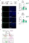
Fig. 8. Selective inhibition of striatal Parvalbumin-positive cells in PV-Cre mice rescues the memory capacity impairment induced by the higher dose of SKF 38393.
a Schematic of the experimental design. PV-Cre mice were infected with AAV-hSyn-DIO-hM4D(Gi)-mCherry in the striatum (STR). Before undergoing the 6 different objects test (6-DOT), mice were treated with SKF 0.1 mg/kg together with vehicle or CNO 1 mg/kg. b The discrimination index in double injected mice (SKF + CNO), but not of the CNO group (vehicle + CNO), was similar to that of the control group (Vehicle + Vehicle) (Two-way ANOVA: Pre-treatment SKF 0.1/ vehicle: F1,22 = 0.07; p = 0.7807; Treatment CNO/ vehicle: F1,22 = 0.5; p = 0.4806; Pre-treatment x Treatment: F1,22 = 15.95; p = 0.0006, Tukey’s post hoc). c Representative images of immunofluorescence staining using an anti-c-Fos antibody and (d) relative quantification. CNO-SKF injected animals show increased c-Fos+ cells in the STR compared to control mice injected with SKF 0.1 or Vehicle (Veh) (Two-way ANOVA: Pre-treatment SKF 0.1/ vehicle: F1,16 = 8.8; p = 0.0090; Treatment CNO/ vehicle: F1,16 = 1.7; p = 0.2007; Pre-treatment x Treatment: F1,16 = 16.26; p = 0.0010, Tukey’s post hoc). Scale bars: 100 µm. e Graphical abstract of the proposed mechanism. The memory enhancing effects of D1 agonists are regulated by the recruitment of cAMP/PKA in the striatum. The memory impairing effects of a high dose of D1 agonists are due to cAMP/PKA activation in the medial prefrontal cortex, which leads to a recruitment of GABA interneurons in the dorsomedial striatum, preventing its activation. Created in BioRender https://BioRender.com/x47z499. Data in bar charts are presented as mean values  ±  SEM. *p < 0.05 vs SKF + Vehicle; #p < 0.05 vs all groups. Source data are provided in the Source Data file.
Fig. 9
Fig. 9. SKF 38393 rescues memory capacity impairment in a pharmacological animal model of schizophrenia.
a Schematic of the experimental design of MK-801 treatment. Mice were treated with vehicle or MK-801 for 7 days, followed by two days of wash-out. They were then tested in the 6 different objects test (6-DOT) or 6 identical objects test (6-IOT). The day after, mice were divided into four sub-groups: vehicle, haloperidol, SKF 38393, and haloperidol plus SKF. The treatment lasted 14 days. On the last day, they were retested in the 6-DOT or 6-IOT under drug. b Effects of sub-chronic treatment of MK-801 (0.3 mg/kg) on the discrimination index during the 6-IOT (One-way ANOVA: Treatment: F1,46 = 0.009; p = 0.9239) and (c) the 6-DOT (One-way ANOVA: Treatment: F1,46 = 24.91; p < 0.0001). MK-801 specifically impaired memory capacity in high but not in low memory load condition. d Effects of haloperidol (Halo) (0.075 mg/kg), SKF 38393 (0.001 mg/kg), and haloperidol in combination with SKF on the discrimination index during the 6-IOT (Two-way ANOVA: Pre-treatment: F1,56 = 0.08; p = 0.7777; Post-treatment: F3,56 = 2.33; p = 0.0832; Pre-treatment x Post-treatment: F3,56 = 0.06; p = 0.9775) and (e) 6-DOT (Two-way ANOVA: Pre-treatment: F1,56 = 4.06; p = 0.0486; Post-treatment: F3,56 = 3.93; p = 0.0128; Pre-treatment x Post-treatment: F3,56 = 2.48; p = 0.0700, Tukey’s post hoc). Haloperidol, when given chronically, dramatically impaired performance in the DOT in both vehicle and MK-801 pretreated animals. SKF rescued memory capacity impairment but did not prevent the impairing effects of haloperidol. Data in bar charts are presented as mean values  ±  SEM. *p < 0.05 vs vehicle. Source data are provided in the Source Data file.

References

    1. Miller, G. A. The magical number seven, plus or minus two: Some limits on our capacity for processing information. Psychol. Rev. 10.1037/0033-295x.101.2.343 (1994). - PubMed
    1. Sannino, S. et al. Role of the dorsal hippocampus in object memory load. Learn. Mem. 10.1101/lm.025213.111 (2012). - PubMed
    1. Assecondi, S., Hu, R., Kroeker, J., Eskes, G. & Shapiro, K. Older adults with lower working memory capacity benefit from transcranial direct current stimulation when combined with working memory training: a preliminary study. Front. Aging Neurosci.14, 1–11 (2022). - PMC - PubMed
    1. Leonard, C. J. et al. Toward the neural mechanisms of reduced working memory capacity in schizophrenia. Cereb. Cortex10.1093/cercor/bhs148 (2013). - PMC - PubMed
    1. Fuster, J. M. & Alexander, G. E. Neuron activity related to short-term memory. Science173, 652–654 (1971). - PubMed

MeSH terms

Substances

LinkOut - more resources