. 2025 Mar 17;16(1):2623.
doi: 10.1038/s41467-025-57969-2.

Genetically encoding ε-N-methacryllysine into proteins in live cells

Affiliations

Genetically encoding ε-N-methacryllysine into proteins in live cells

Tian-Yi Zhu et al. Nat Commun. .

Abstract

Lysine acylation is a ubiquitous post-translational modification (PTM) that plays pivotal roles in various cellular processes, such as transcription, metabolism, protein localization and folding. Thousands of lysine acylation sites have been identified based on advances in antibody enrichment strategies, highly sensitive analysis by mass spectrometry (MS), and bioinformatics. However, only 27 lysine methacrylation (Kmea) sites have been identified exclusively in histone proteins. It is hard to separate, purify and differentiate the Kmea modification from its structural isomer lysine crotonylation (Kcr) using general biochemical approaches. Here, we identify Kmea sites on a non-histone protein, Cyclophillin A (CypA). To investigate the functions of Kmea in CypA, we develop a general genetic code expansion approach to incorporate a non-canonical amino acid (ncAA) ε-N-Methacryllysine (MeaK) into target proteins and identify interacting proteins of methacrylated CypA using affinity-purification MS. We find that Kmea at CypA site 125 regulates cellular redox homeostasis, and HDAC1 is the regulator of Kmea on CypA. Moreover, we discover that genetically encode Kmea can be further methylated to ε-N-methyl-ε-N-methacrylation (Kmemea) in live cells.


Conflict of interest statement

Competing interests: The authors declare no competing interests.

Figures

Fig. 1
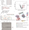
Fig. 1. Identification of Kmeas on CypA protein.
a Tandem mass spectrum of BVSB mediated cross-linked peptide between TXN1 and CypA. b Previously, protein interaction of TXN1 and CypA was identified in TXN1 His-tag pull down. To validate protein interaction of TXN1 and CypA, CypA IP experiment was performed and TXN1 was identified in CypA IP samples. c, d HEK293T cells were transfected with CpyA-Strep, Strep-tag purified CypA was digested with trypsin. Digested peptides were analyzed by mass spectrometry. Tandem mass spectrum of peptides contained methacrylation on CypA Lys131 (c) and Lys125 (d). The K in red represents the Kmea modification. e Validation of Kmea on CypA by Western blot analysis. When CypA Lys125 was mutated to Arg, the Kmea level of CypA dramatically decreased. The experiment was repeated three times with similar results. When CypA Lys131 was mutated to Arg, the Kmea level of CypA partially decreased. f Proposed fragmentation pathway of CycIm ion from methacrylated peptide. g CycIm ion was mapped on MS/MS spectrum of methacrylated peptide (Kmea on CypA Lys131). h Confirm CycIm ion with chemically synthesized standard methacrylated peptide.
Fig. 2
Fig. 2. Genetically encode MeaK into proteins in E. coli and mammalian cells.
a Chemical structure of Meak. b Left: X-ray crystal structure of the MmPylRS complex with pyrrolysyl-AMP (PDB ID: 2Q7H), active-site residues for mutation are highlighted in green. Right: residue numbering is based on MbPylRS and MmPylRS. c SDS-PAGE gel of His-tag purified MBP-Z E24MeaK. Protein Z is IgG Fc-binding domain, MBP-Z is maltose binding protein tagged protein Z. d Intact protein mass analysis of purified protein from E. coli expressed MBP-Z E24MeaK. e Tandem mass spectrum of MeaK incorporated peptide of EGFP D190MeaK. The red U represents MeaK incorporation site. f CycIm ion was mapped on the MS/MS spectrum of the MeaK incorporated peptide. g Genetically encode Meak into EGFP protein in mammalian cells. FACS analysis of HEK293T cells after MeaK incorporation into EGFP-151TAG. h Left: Fluorescence images of EGFP Y151MeaK showing incorporation of MeaK into EGFP in HEK293T cells. Right: Select images from different fields of view under a 10× objective lens, and use ImageJ software to calculate the mean fluorescence intensity (n = 5). Error bar represents ± one standard deviation (SD). i Intact protein mass analysis of HEK293T cells expressed EGFP Y151MeaK. j Tandem mass spectrum of MeaK incorporated peptide of EGFP Y151MeaK.
Fig. 3
Fig. 3. Genetically encode MeaK into CypA to identify interacting proteins of methacrylated CypA.
a Alignment of K125 and K131-flanking sequences of CypA from multiple vertebrate species. CypA K125 and CypA K131 are highlighted in red. b Comparison of CypA K125 acetylation levels between tumor and adjacent tissues from different cancer types. Raw data were obtained from six acylated proteomes of CPTAC (Clinical Proteomic Tumor Analysis Consortium) database. **P < 0.01 (two-tailed t-test). p = 0.0228 and 0.0046, respectively. The center line denotes the median value, the box contains the 25th to 75th percentiles. The whiskers mark the minima and maxima. Sample numbers are provided in the Source Data file. c Tandem mass spectrum of MeaK incorporated peptide from HEK293T cells expressed CypA K125MeaK. The U in red contains the Kmea modification. d SDS-PAGE gel analysis of CypA (K125MeaK, K125A, WT) IP with three biological replicates. Nanobody comes from alpaca-derived heavy chain antibody fragment (VHH) carried by Strep beads. e Valcano plot of semi-quantification result on CypA and MeaK incorporated CypA IP. Red dots, significantly enriched hits (Fold Change ≥2, p-value < 0.05). Gray dots, depleted hits or non-differentially expressed proteins. Data quantified with the Empirical Bayes test two-sided. f Biological process analysis of the enriched hits in (e) and unique proteins identified in CypA K125MeaK in Strep-IP.
Fig. 4
Fig. 4. Kmea at site 125 of CypA increased ROS level in HeLa cells.
ac HeLa cells were transfected with CypA-WT-mCherry or CypA-K125MeaK-mCherry, which were either treated with/without 4 µM CsA for 36 h. ROS was measured by flow cytometry. Fluorescence intensity of FITC channels was detected after selection of the same number of CypA+ cells by ECD channel. d Quantitative comparison of ROS levels in different cells. n = 3 independent experiments. ns, not significant; *p < 0.05; **p < 0.01, p = 0.0402 and 0.0048, respectively; (two-tailed t-test/One-way ANOVA analysis). Error bar represents ±one standard deviation. e Model showing CypA 125Kmea modification increases ROS level of cells and it blocks the suppression of CsA on CypA.
Fig. 5
Fig. 5. Identification of erasers of Kmea on CypA.
a Genetically encoded EGFP K85TAG-based probe for de-conjucation enzymes of Kmea. b Top: Construct of Fluorescent reporter containing EGFP K85TAG, self-cleavable peptide P2A and mCherry. Down: Fluorescence imaging of HDACs demethacrylation activity in HEK293T cells. The cells were co-transfected with two plasmids: one containing chPylRS Y384F/tRNAPylCUA and another containing mCherry-EGFP K85TAG; MeaK (final concentration 1 mM) and NaBu (final concentration 10 mM) were added after transfection 6 h. Imaging was carried out after 36 h. Scale bars, 150 µm. c Normalizing signal of EGFP with mCherry. The error bars represent standard deviation from the mean. n = 3 independent experiments. ns, not significant; ***p < 0.001, p = 0.0004. d Binding of deacetylases to methacrylated CypA, which were detected by AP-MS from HEK293T cells expressed CypA K125MeaK. e Interactions between methacrylated CypA and HDACs with Co-IP experiments. f Western blot analysis showing successful demethacrylation assay by HDAC1 in vivo. g Purificated CypA K125MeaK protein was incubated with or without recombinant HDAC1 for 2 h at 30 °C. Samples were then analyzed by Western blot.
Fig. 6
Fig. 6. Methylation on genetically encoded Kmea in live cells.
a Chemical structure of Kacme. b Chemical structure of Kmemea. c Tandem mass spectrum of methylation on CypA K125MeaK. d Tandem mass spectrum of methylation on CypA K131MeaK. e Proposed fragmentation pathway of LinIm ion from methyl-methacrylated peptide. f LinIm ion on tandem mass spectrum of peptide containing CypA K125Kmemea. g LinIm ion on tandem mass spectrum of peptide containing CypA K131Kmemea.

References

    1. Shang, S., Liu, J. & Hua, F. Protein acylation: mechanisms, biological functions and therapeutic targets. Signal Transduct. Target. Ther.7, 396 (2022). - PMC - PubMed
    1. West, A. C. & Johnstone, R. W. New and emerging HDAC inhibitors for cancer treatment. J. Clin. Investig.124, 30–39 (2014). - PMC - PubMed
    1. Xie, Z. et al. Metabolic regulation of gene expression by histone lysine beta-hydroxybutyrylation. Mol. cell62, 194–206 (2016). - PMC - PubMed
    1. Tian, H. et al. Genetically encoded benzoyllysines serve as versatile probes for interrogating histone benzoylation and interactions in living cells. ACS Chem. Biol.16, 2560–2569 (2021). - PubMed
    1. Zhang, D. et al. Metabolic regulation of gene expression by histone lactylation. Nature574, 575–580 (2019). - PMC - PubMed

LinkOut - more resources