. 2025 Mar 17;8(1):448.
doi: 10.1038/s42003-025-07697-6.

Smooth muscle cell Piezo1 depletion results in impaired contractile properties in murine small bowel

Affiliations

Smooth muscle cell Piezo1 depletion results in impaired contractile properties in murine small bowel

Geoanna M Bautista et al. Commun Biol. .

Abstract

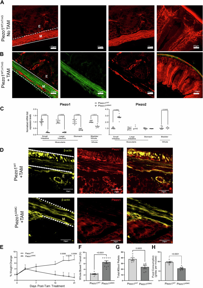
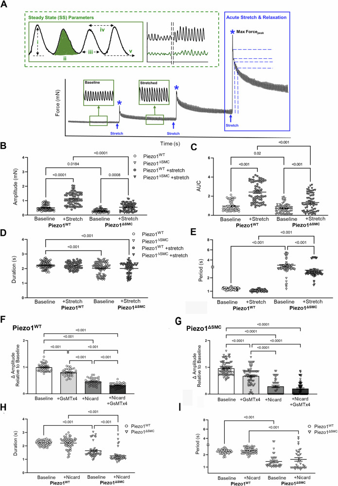
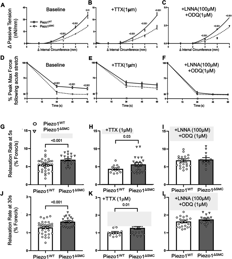
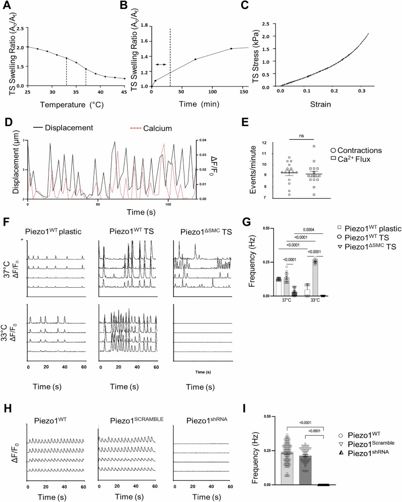
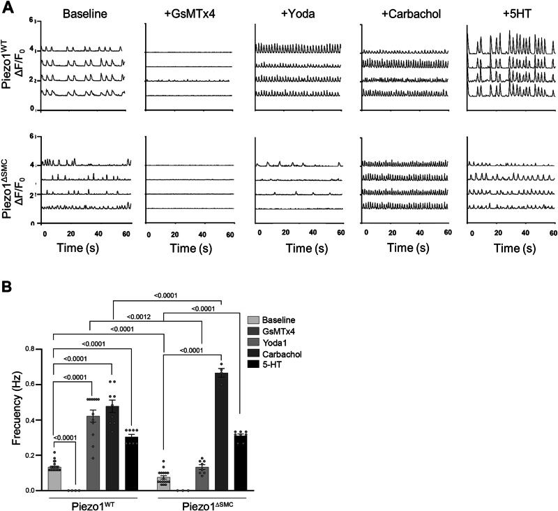
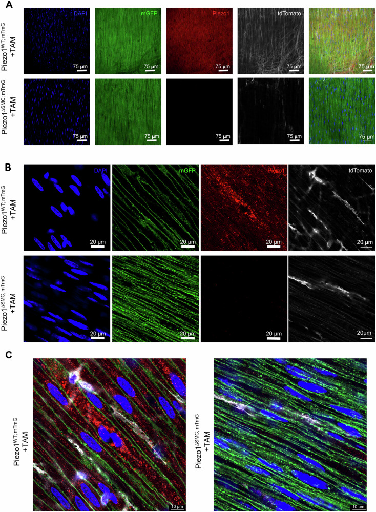
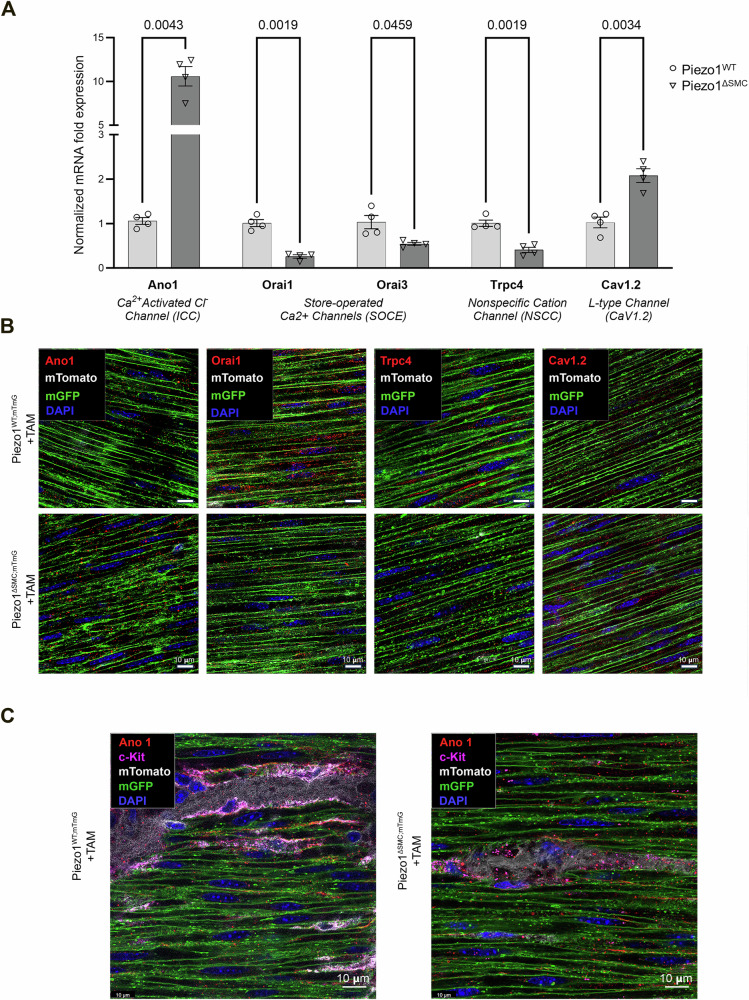
Piezo1 is a mechanosensitive cation channel expressed in intestinal muscularis cells (IMCs), including smooth muscle cells (SMCs), interstitial cells of Cajal, and Pdgfrα+ cells, which form the SIP syncytium, crucial for GI contractility. Here, we investigate the effects of SMC-specific Piezo1 deletion on small bowel function. Piezo1 depletion results in weight loss, delayed GI transit, muscularis thinning, and decreased SMCs. Ex vivo analyses demonstrated impaired contractile strength and tone, while in vitro studies using IMC co-cultures show dysrhythmic Ca2+ flux with decreased frequency. Imaging reveal that Piezo1 localizes intracellularly, thereby likely impacting Ca2+ signaling mechanisms modulated by Ca2 + -handling channels located on the sarcoplasmic reticulum and plasma membrane. Our findings suggest that Piezo1 in small bowel SMCs contributes to contractility by maintaining intracellular Ca2+ activity and subsequent signaling within the SIP syncytium. These findings provide new insights into the complex role of Piezo1 in small bowel SMCs and its implications for GI motility.


Conflict of interest statement

Competing interests: None of the authors have any potential financial, professional, or personal conflicts.

Figures

Fig. 1
Fig. 1. Inducible SMC-specific Piezo1 depletion results in poor growth and delayed bowel transit.
Efficient Tam-induced translocation of Cre-ER into the nucleus was confirmed in distal small bowel segments taken from 7-9 week-old mice following Tam administration at 4-6 weeks old, with all assessments performed age-matched in both groups. A Myh11-Cre expression in the distal small bowel segment from Piezo1WT mice with membrane-associated Tdtomato fluorescence (mTmG) reporter (Piezo1WT;mTmG) shows exclusive Tdtomato fluorescence in membranes of all cell types across the layers of the bowel in transverse-sections of WM preparations, indicating an absence of Cre expression or leakage without Tam induction. B In contrast, similarly aged mice post-Tam treated Piezo1WT;mTmG mice, the epithelial layer retained Tdtomato, while the cellular components of the muscularis were either Tdtomato or GFP + . Leica Confocal SP8-STED microscope with scale bars set at 10, 20, and 40 μm. C mRNA expression of Piezo1 and Piezo2 in Piezo1ΔSMC and Piezo1WT mice from muscularis samples isolated from the distal small intestine, proximal colon and stomach, and full-thickness samples from the bladder. qPCR was performed using the ΔΔCT method relative to Gapdh expression. Multiple t-test analyses with Welch’s correction were performed with a False Discovery Rate (FDR) set to 5% with the corrected p-values as indicated above comparison groups. Data are shown as mean ± SEM, n = 4–6 mice per group. D Frozen transverse-section of distal small bowel from Piezo1WT (upper) and Piezo1ΔSMC (lower) mice post-Tam, stained with anti-β-actin (yellow) and Piezo1 (Alamone Labs; red) antibodies and imaged with a Zeiss Confocal LSM880 at 40× magnification with 20 µm scale bars. The parallel dashed lines indicate the location of the muscularis (M). Beneath the bottom line is the epithelial (E) layer. E Body weight at various days post-Tam treatment in Piezo1WT and Piezo1ΔSMC mice. F Carmine-red dye was given via oral gavage, and the time to the first red stool pellet was recorded to indicate whole bowel transit time in both groups of mice at 7–9 weeks old following tamoxifen administration at 4-6 weeks old. Total number of hours capped at 8 h. G A total number of stool pellets was produced after 8 h in both groups of mice. H Daily food consumption in both groups of mice. Two-tailed t-test with Welch’s correction performed with a significance set at p < 0.05 is shown above. Data are shown as mean ± SEM, N = 6–10 mice per group.
Fig. 2
Fig. 2. Impaired contractile properties with loss of Piezo1 in SMCs.
Isotonic force measurements were obtained from distal ileal ring segments (3 mm long) from 4- to 6-week-old mice at least 21 days post-Tam at baseline and following acute stretch using a multi-wire myograph. A General schematic providing an overview of the experimental approach. Green dotted box illustrating assessments performed at SS (green) or once the contractions have plateaued following stretch: i. amplitude (mN), the active or contractile force (height of contraction from passive force (v)), ii. AUC (mN-s) measuring the work, iii. duration (s), or time of the entire contraction, iv. period (s) or time between peaks was used to quantify dysrhythmic patterns observed during trials. Representative tracings of Piezo1WT (black) and Piezo1ΔSMC (green). Phasic contractile activity, including (B) amplitude, (C) AUC, (D) duration, and (E) period, were altered in Piezo1ΔSMC mice compared to controls (n = 50 samples, N = 3 mice per group). The effects of GsMTx4 and Nicardipine inhibitor exposure on the contraction amplitudes for (F) Piezo1WT and (G) Piezo1ΔSMC groups. H Duration and (I) period changes in contraction behavior when Piezo1WT and Piezo1ΔSMC are only exposed to nicardipine. For F–I, n ≥ 50 data points from N = 3 mice per group were analyzed with one-way ANOVA with multiple comparison tests.
Fig. 3
Fig. 3. SMC-specific Piezo1 depletion alters tonicity and tissue structure.
Length-tension relationships were assessed using passive tension following stretch at increasing circumferential stretches to determine tissue tonicity. Linear regression analysis with multiple t-tests showed (A) a reduction in the length-tension curve in Piezo1ΔSMC bowel segments, which persisted in the presence of (B) TTX and (C) L-NNA/ODQ. Viscoelastic properties were assessed using percent reduction from max (peak) force (Fig. 2A, blue asterisks) over time until SS was reached. Compared to Piezo1WT, Piezo1ΔSMC bowel segments had (D) a greater reduction in max (peak) force at baseline conditions than in the presence of (E) TTX and (F) L-NNA/ODQ. (N ≥ 4 mice per group, analyzed with simple linear regression and multiple t-tests with Bonferroni correction). The rate of relaxation from the max force was increased in Piezo1ΔSMC bowel at (G) 5 s and (J) 30 s, which was retained with the addition of (H, K) TTX but not with (I, L) L-NNA/ODQ. (N = 3 mice per group were analyzed with Welch’s t-test with multiple comparison tests.
Fig. 4
Fig. 4. Thermosensitive stretch-inducible hydrogels with co-cultured IMCs showed altered Ca2+ signaling with loss of SMC Piezo1.
A Hydrogel properties showing a temperature range of 37 °C–33 °C selected for TS scaffolds to achieve optimal surface area change based on equilibrium swelling behavior. B TS scaffold swelling rate/ratio determined the time required to achieve stretching of IMCs in an isotropic manner, with a dashed line to mark the operation range of 30 min for a 20% area increase. C The stress-strain behavior of TS scaffolds (4.1 kPA) was modified to simulate the mechanical properties of small intestine tissue (4.1–4.5 kPA). D IMCs isolated from external muscularis strips from 8 to 10-day-old murine pups (N = 6–8 murine pups per biological sample) seeded on plastic or thermosensitive (TS) stretch-inducible hydrogels with spontaneous contractile behavior. Representative tracings depicting contractions (black) as measured by displacement (left axis) overlapped Ca2+ flux (red dashed) measured by absolute intensity changes, ΔF/F0 (right axis) with (E) frequency measurements of contractions and Ca2+ flux shown (n > 12–15 time points per group). F Representative GCaMP6f Ca2+ tracings (ΔF/F0) of PiezoWT IMCs seeded on plastic and TS hydrogels that stretch with a temperature reduction from 37 °C to 33 °C compared to Piezo1ΔSMC IMCs seeded on TS. G Two-way ANOVA was used to measure change in frequency with change in temperature using Sidak correction. Significance values indicated in figure. Data displayed as mean ± SEM, n > 6 biological samples for each group (5–8 murine pups per biological sample). H IMCs isolated from muscularis layers of murine 10-day-old pups (N = 6–8 murine pups per biological sample). Representative GCaMP6f Ca2+ tracings (ΔF/F0) of Piezo1WT, Piezo1SCRAMBLE, and Piezo1shRNA IMCs seeded on a plastic scaffold at baseline, measuring (I) frequency differences (n > 10 biological samples per group, N = 6–8 murine pups per biological sample).
Fig. 5
Fig. 5. Piezo1 modulators on in vitro Ca2+ flux.
IMCs were isolated from muscularis layers of murine 8-10-day-old pups (N = 6–8 murine pups per biological sample). A Representative GCaMP6f Ca2+ tracings (ΔF/F0) of Piezo1WT and Piezo1ΔSMC IMCs with GsMTx4 (20 µM), Yoda1 (5 µM), Carbachol (10 µM) and 5-HT (10 µM) between Piezo1WT and Piezo1ΔSMC with (B) frequency differences between groups (n > 6 biological samples per group, N = 6–8 murine pups per biological sample) compared. Two-way ANOVA with interaction followed by Tukey’s post-hoc analysis was applied.
Fig. 6
Fig. 6. Cellular composition of the muscularis layer following the loss of SMC Piezo1.
A Total muscularis thickness was assessed on H&E (right) stained FFPE samples from distal small bowel segments of Piezo1WT and Piezo1ΔSMC mice at 7–10 weeks old following post-Tam administration performed at 4–6 weeks old. 8–10 representative regions per mouse were measured from 3 to 4 mice per group. B The SMC density (SMCs per mm2) in circular and longitudinal layers in a WM preparation is decreased in Piezo1ΔSMC distal small bowel compared to Piezo1WT mice (N = 3–4 mice per group). C–E ICC, Pdgfrα + , and glial cell density as measured by staining for c-Kit (R&D Systems), Pdgfrα (Cell Signaling), and Gfap (Abcam) of WM preparations in Piezo1ΔSMC mice compared to Piezo1WT. Neuronal number of (F) cells, (G) fibers, and (H) bundle density per mm2 as measured by staining for Tubb3 (Abcam) in Piezo1ΔSMC mice compared to Piezo1WT using WM preparations. Unpaired two-tailed t-test with Welch’s correction. Data presented as mean ± SEM, n > 18 areas (N = 3–4 mice/group) with p-values indicated above, significance set at p < 0.05.
Fig. 7
Fig. 7. Expression and localization of Piezo1 in small bowel muscularis.
A–C WM prepared distal bowel samples from Piezo1WT;mTmG and Piezo1ΔSMC;mTmG mice and 21 d post-Tam were used for IF staining of Piezo1 as imaged by Leica Stellaris 8 microscope (A), and Leica Confocal SP8-STED microscope (B, C). Green shows the membrane-GFP; pseudo-white is the membrane-Tdtomato from the mTmG reporter, and the anti-Piezo1 (Alamone Labs) signal is pseudo-red. The scale bars are (A), 75 μm; (B), 20 μm; and (C) 10 μm, respectively.
Fig. 8
Fig. 8. SMC-specific deletion of Piezo1 alters related ion channel expression within the muscularis.
A Transcriptional expression (mRNA) of Ano1, Orai1, Orai3, Trpc4, and Cav1.2 in Piezo1ΔSMC and Piezo1WT mice from muscularis samples isolated from the distal small intestine. Normalized mRNA fold expression based on qRT-PCR results was calculated using the ΔΔCT method relative to Gapdh expression. Multiple t-test analyses with Welch’s correction were performed with a False Discovery Rate (FDR) set to 5% with the corrected p-values (q-values) above comparison groups. Data are shown as the mean ± SEM (N  = 4–5 mice per group). B WM distal small bowel samples from Piezo1WT;mTmG and Piezo1ΔSMC;mTmG mice 21 d post-Tam were used for IF staining with a Leica Confocal SP8-STED microscope of various proteins required for Ca+2 homeostasis. Green shows the membrane-GFP; pseudo-white is the membrane-Tdtomato from the mTmG reporter. Antibodies against Orai1, Trpc4, Cav1.2, and Ano1 (Alomone Labs) were pseudo-red. Scale bars, 10 μm. C Imaging of the muscularis from WM-prepared samples stained for Ano1 and c-Kit indicates Ano1 co-localization with c-Kit as expected in Piezo1WT;mTmG sample, and an increase of Ano1 staining along the GFP+ plasma membrane of SMCs in Piezo1ΔSMC;mTmG mice.
Fig. 9
Fig. 9. Piezo1 protein in SMCs colocalizes with IP3R and RyR.
WM sample of distal small bowel isolated from Piezo1WT;mTmG and Piezo1ΔSMC;mTmG mice 21 d post-Tam, were imaged with Leica Confocal SP8-STED microscope. A The anti-RyR (Santa Cruz) signal is pseudo-yellow, the anti-Piezo1 (Alamone Labs) signal is pseudo-red, green shows the membrane-GFP, and pseudo-white is the membrane-Tdtomato from the mTmG reporter. All scale bars are at 10 μm and insert at 5 μm. B The anti-IP3R (BD Bioscience) signal is pseudo-yellow, while the anti-Piezo1 antibody and other signals from the mTmG reporter are identical to A. Immunofluorescence-stained IP3 and RYR protein channels were quantified using AIVIA software Pixel Classifier and Smart Segmentation tools to segment structures, distinguish them from the background, and generate 2D outlines. Total counts were then normalized by ROI area relative to sample data to account for batch and animal differences (n = 25 ROIs per mouse, N = 3 mice per group). Data shown as mean +/− SEM. Statistical analysis was performed on log-transformed averages per mouse per group with significant p-values indicated above, with full nested analysis included in supplementary fig. 8. C, D Isometric force assessments on intact, full-thickness bowel segments from Piezo1ΔSMC mice have impaired response to carbachol at SS compared to Piezo1WT, including amplitude, (D) period, and (E) duration (n ≥ 20 measurements from N = 3 mice per group).

References

    1. Mazet, B. Gastrointestinal motility and its enteric actors in mechanosensitivity: past and present. Pflügers Archiv: Eur. J. Physiol.467,191–200 (2014). - PubMed
    1. Furness, J. B, Kunze, W. A & Clerc, N. Nutrient tasting and signaling mechanisms in the gut. II. The intestine as a sensory organ: neural, endocrine, and immune responses. Am. J. Physiol.277, G922–G928 (1999). - PubMed
    1. Holm, A. N., Rich, A., Sarr, M. G & Farrugia, G. Whole cell current and membrane potential regulation by a human smooth muscle mechanosensitive calcium channel. Am. J. Physiol. Gastrointest. Liver Physiol.279, G1155–G1161 (2000). - PubMed
    1. Bertrand, P. P. Real-time measurement of serotonin release and motility in guinea pig ileum. J. Physiol.577, 689–704 (2006). - PMC - PubMed
    1. Costa, M. & Furness, J. B The peristaltic reflex: an analysis of the nerve pathways and their pharmacology. Naunyn Schmiedebergs Arch. Pharm.294, 47–60 (1976). - PubMed