Review
. 2025 Mar 17;23(1):341.
doi: 10.1186/s12967-025-06291-y.

New insights on anti-tumor immunity of CD8+ T cells: cancer stem cells, tumor immune microenvironment and immunotherapy

Affiliations
Review

New insights on anti-tumor immunity of CD8+ T cells: cancer stem cells, tumor immune microenvironment and immunotherapy

Yibin Lin et al. J Transl Med. .

Abstract

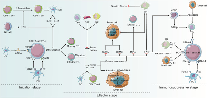
Recent breakthroughs in tumor immunotherapy have confirmed the capacity of the immune system to fight several cancers. The effective means of treating cancer involves accelerating the death of tumor cells and improving patient immunity. Dynamic changes in the tumor immune microenvironment alter the actual effects of anti-tumor drug production and may trigger favorable or unfavorable immune responses by modulating tumor-infiltrating lymphocytes. Notably, CD8+ T cells are one of the primary tumor-infiltrating immune cells that provide anti-tumor response. Tumor cells and tumor stem cells will resist or evade destruction through various mechanisms as CD8+ T cells exert their anti-tumor function. This paper reviews the research on the regulation of tumor development and prognosis by cancer stem cells that directly or indirectly alter the role of tumor-infiltrating CD8+ T cells. We also discuss related immunotherapy strategies.

Keywords: Cancer prognosis; Cancer stem cell (CSC); Cytotoxic CD8+ T lymphocyte (CTL); Immunotherapy; Tumor immune microenvironment (TIME).


Conflict of interest statement

Declarations. Ethics approval and consent to participate: Not applicable. Consent for publication: Not applicable. Competing interests: The authors declare that they have no competing interests.

Figures

Fig. 1
Fig. 1
The process of CD8+ T cell infiltration into tumor tissue Tumor cells release tumor antigens (Tas) into the tumor microenvironment (TME), which include tumor-associated antigens (TAAs) and tumor-specific antigens (TSAs). In lymph nodes, antigen-presenting cells (APCs), such as dendritic cells (DCs), identify and bind to TAs. Tumor-infiltrating APCs carrying TAs then migrate to the lymph node, process and present TAs to CD8+ T cells, activating them and producing TA-specific CD8+ T cells. CD49a and CD103 are expressed on the surface of TA-specific CD8+ T cells and can bind to E-cadherin on tumor cells. TA-specific CD8+ T lymphocytes infiltrate tumor tissue, recognize TAs on tumor cells, and induce tumor cell death
Fig. 2
Fig. 2
Activation and deactivation of CD8+ T cells Dendritic cells (DCs), natural killer (NK) cells, and CD4+ T cells play significant roles in the activation of CD8+ T cells. DCs interact with CD8+ T lymphocytes via receptor ligands. CD4+ T cells facilitates the activation of CD8+ T cells by stimulating them. DCs promote the development of CD4+ T cells into antigenspecific effector T cells. CD4+ T cells stimulate CD8+ T cell start via cytokines. CD4+ T cells can also contribute to DC activation and licensing by inducing DC maturation, co-stimulatory molecule expression, and cytokine secretion, all of which activate CD8+ T cells. NK cells also perform similar functions. In the effector stage, CTLs are activated to kill target cells by granule cytotoxicity and Fas ligand (FasL)-mediated apoptosis. CTLs emit IFN-γ and TNF-α, causing cytotoxicity in cancer cells. NK cells play similar functions. In the immunosuppressive phase, activated CTLs are activated and licensed to express co-stimulatory molecules and secrete cytokines. In the immunosuppressive stage, activated T cells begin to express co-inhibitory receptors, such as the programmed death-1 receptor (PD-1), within hours or days of activation. This occurs through IFN-γ induction of programmed death- 1 ligand (PD-L1) expression in anti-tumor M1 macrophages and cancer cells. Expression of CTL-associated antigen 4 (CTLA-4) by regulatory T cells (Tregs) can also inhibit the suppressive activity of CD8+ T cells, thereby triggering immunosuppressive activity within the TME
Fig. 3
Fig. 3
Interactions between cancer stem cells (CSCs) and immune cells in the tumor immune microenvironment (TIME) CSCs enhance the activity of type 2 macrophages (M2), type 2 neutrophils (N2), and myeloid-derived suppressor cells (MDSCs) by producing the transforming growth factor-β (TGF-β), thereby promoting the epithelial-mesenchymal transition (EMT) of tumor cells. EMT is positively correlated to the expression of programmed death ligand 1 (PD-L1) on M2 macrophages and tumor cells, which damages the function of CD8+ T cells by increasing the activity of PD-1 and stimulating the release of vascular endothelial growth factor (VEGF). Moreover, CSCs generate immunosuppressive factors such as IL-4, IL-10, and IL-13 to prevent the maturation of dendritic cells (DCs), which weakens the activation of CD8+ TILs
Fig. 4
Fig. 4
T cell dependent cancer immunotherapy Effective strategies to enhance CD8+ T cell infiltration and effector cell function include autologous back-treatment with chimeric antigen receptor (CAR) T cells, TCR-transduced T cells, and tumor-infiltrating T cells (TILs). Inhibiting antibodies can also be used to suppress the T cell co-inhibitory axis (e.g., B7/CTLA-4). Antagonistic antibodies can induce T cell co-stimulatory axis (e.g., 4-1BB/4-1BBL)

References

    1. Wherry EJ, Ha S-J, Kaech SM, Haining WN, Sarkar S, Kalia V, et al. Molecular signature of CD8 + T cell exhaustion during chronic viral infection. Immunity. 2007;27:670–84. - PubMed
    1. Barber DL, Wherry EJ, Masopust D, Zhu B, Allison JP, Sharpe AH, et al. Restoring function in exhausted CD8 T cells during chronic viral infection. Nature. 2006;439:682–7. - PubMed
    1. Lapidot T, Sirard C, Vormoor J, Murdoch B, Hoang T, Caceres-Cortes J, et al. A cell initiating human acute myeloid leukaemia after transplantation into SCID mice. Nature. 1994;367:645–8. - PubMed
    1. Bonnet D, Dick JE. Human acute myeloid leukemia is organized as a hierarchy that originates from a primitive hematopoietic cell. Nat Med. 1997;3:730–7. - PubMed
    1. Prager BC, Xie Q, Bao S, Rich JN. Cancer stem cells: the architects of the tumor ecosystem. Cell Stem Cell. 2019;24:41–53. - PMC - PubMed