. 2025 Mar 18:13:RP93754.
doi: 10.7554/eLife.93754.

Pain persists in mice lacking both Substance P and CGRPα signaling

Affiliations

Pain persists in mice lacking both Substance P and CGRPα signaling

Donald Iain MacDonald et al. Elife. .

Abstract

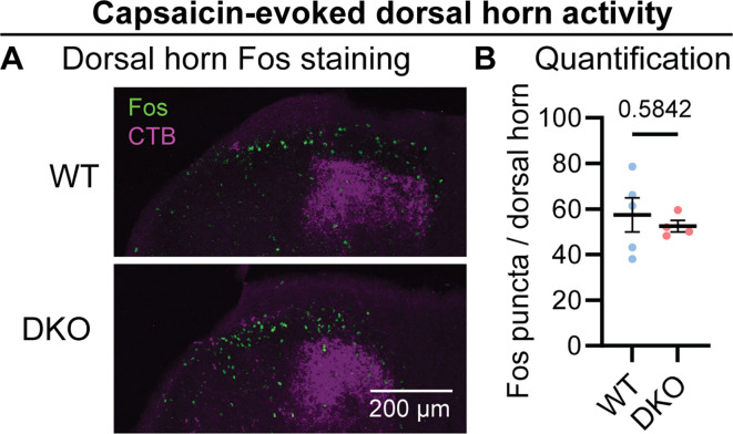
The neuropeptides Substance P and CGRPα have long been thought important for pain sensation. Both peptides and their receptors are expressed at high levels in pain-responsive neurons from the periphery to the brain making them attractive therapeutic targets. However, drugs targeting these pathways individually did not relieve pain in clinical trials. Since Substance P and CGRPα are extensively co-expressed, we hypothesized that their simultaneous inhibition would be required for effective analgesia. We therefore generated Tac1 and Calca double knockout (DKO) mice and assessed their behavior using a wide range of pain-relevant assays. As expected, Substance P and CGRPα peptides were undetectable throughout the nervous system of DKO mice. To our surprise, these animals displayed largely intact responses to mechanical, thermal, chemical, and visceral pain stimuli, as well as itch. Moreover, chronic inflammatory pain and neurogenic inflammation were unaffected by loss of the two peptides. Finally, neuropathic pain evoked by nerve injury or chemotherapy treatment was also preserved in peptide-deficient mice. Thus, our results demonstrate that even in combination, Substance P and CGRPα are not required for the transmission of acute and chronic pain.

Keywords: mouse; neuropeptides; neuroscience; pain; somatosensation.


Conflict of interest statement

DM, MJ, JS, RB, AN No competing interests declared, AC Reviewing editor, eLife

Figures

Figure 1.
Figure 1.. Tac1::Calca DKO mice lack Substance P and CGRPα peptides throughout the nervous system.
(A) Confocal images showing Substance P immunostaining in the dorsal root ganglion (DRG), spinal cord dorsal horn and midbrain of a WT mouse (top). No staining is detectable in the DKO (bottom). (B) Confocal images showing CGRP immunostaining in the DRG, spinal cord dorsal horn, and amygdala of a WT mouse (top). The DKO mice show no obvious staining (bottom). Images in both (A) and (B) are representative of staining performed on tissue from 1 M and 1 F animal per genotype. (C) Schematic showing co-culture of Tac1-RFP-labeled DRG neurons and Substance P-sniffer HEK293 cells expressing NKR1, GCaMP6s, and Gαo. Capsaicin-evoked secretion of Substance P from DRG activates the NKR1 receptor in neighboring HEK leading to calcium release from stores and GCaMP6s-mediated fluorescence increase. (D) Fluorescence images showing Tac1-RFP-labeled DRG neurons (magenta) and Substance P-sniffer cells (green). Capsaicin causes an increase in Substance P-sniffer GCaMP6s fluorescence when cultured with DRGs from WT, but not DKO, mice. Application of exogenous Substance P (10 nM) activates Substance P-sniffers in both conditions. (E) Quantification of fluorescence change in Substance P-sniffer cells in response to vehicle, capsaicin, and Substance P stimulation in WT and DKO mice. n=12 wells from 2 mice (1 M, 1 F) for WT, and n=12 wells from 2 mice (1 M, 1 F) for DKO. For (E), means were compared using repeated measures 2-way ANOVA, followed by post-hoc Sidak’s test. Error bars denote standard error of the mean. (F) Schematic showing co-culture of Tac1-RFP-labeled DRG neurons and Substance P-sniffer HEK293 cells expressing Calcrl, Ramp1, GCaMP6s, and Gαo. Capsaicin-evoked secretion of CGRPα from DRG activates the CGRP receptor complex in neighboring HEK leading to calcium release from stores and GCaMP6s-mediated fluorescence increase. (G) Fluorescence images showing Tac1-RFP-labeled DRG neurons (magenta) and Substance P-sniffer cells (green). Capsaicin causes an increase in CGRP-sniffer GCaMP6s fluorescence when cultured with DRGs from WT, but not DKO, mice. Application of exogenous CGRPα at a saturating concentration (100–1000 nM) activates CGRP-sniffers in both conditions. (H) Quantification of fluorescence change in CGRP-sniffer cells in response to vehicle, capsaicin, and CGRPα stimulation in WT and DKO mice. n=18 wells from 3 mice (2 M, 1 F) for WT, and n=18 wells from 3 mice (2 M, 1 F) for DKO. For (H), means were compared using repeated measures two-way ANOVA, followed by post-hoc Sidak’s test. Error bars denote standard error of the mean.
Figure 1—figure supplement 1.
Figure 1—figure supplement 1.. Substance P-sniffer cells selectively respond to Substance P.
(A) Dose-response curve showing Substance P activates Substance P-sniffer cells at low nanomolar concentrations (EC50=11.8 nM), but cells are insensitive to CGRPα. (B) Dose-response curve showing CGRPα activates CGRP-sniffer cells at low nanomolar concentrations (EC50=11.8 nM), but cells are insensitive to Substance P. Four-parameter variable slope dose-response curves were fit by non-linear regression. At least three replicates were performed for each concentration per condition, in two independent experiments.
Figure 2.
Figure 2.. Tac1::Calca DKO mice respond to acute painful and itching stimuli.
(A) 50% withdrawal threshold for von Frey punctate stimulation (log g). n=14 (7M, 7 F) for WT & n=14 (7M, 7 F) for DKO. (B) Percentage response to noxious pinprick stimulation. n=13 (5M, 8 F) for WT and n=11 (4M, 7 F) for DKO. (C) Time spent attending to alligator clip applied to paw for 60 s. n=7 (3M, 4 F) for WT and n=10 (6M, 4 F) for DKO. (D) Latency to withdraw to low and high radiant heat. For low setting, n=12 (6M, 6 F) for WT and n=8 (4M, 4 F) for DKO. For high setting, n=15 (8M, 7 F) for WT and n=17 (9M, 8 F) for DKO. (E) Latency to lick hindpaw following exposure to hot plate at two different temperatures. For 52.5 °C, n=15 (7M, 8 F) for WT and n=10 (6M, 4 F) for DKO. For 55.5 °C, n=9 (6M, 3 F) for WT and n=8 (5M, 3 F) for DKO. (F) Time spent licking in 60 s immediately following acetone application to paw. n=12 (5M, 7 F) for WT and n=12 (5M, 7 F) for DKO. (D) Latency to respond to dry ice. n=19 (8M, 11 F) for WT and n=13 (5M, 8 F) for DKO. (H) Time spent licking in 5 min after capsaicin injection to paw. n=9 (4M, 5 F) for WT and n=8 (4M, 4 F) for DKO. (I) Time spent licking in 5 min after 1% AITC injection to paw. n=12 (6M, 6 F) for WT and n=12 (6M, 6 F) for DKO. (J) Number of writhes in 15 min starting 5 min after intraperitoneal injection of 0.6% acetic acid. n=9 (5M, 4 F) for WT and n=7 (5M, 2 F) for DKO. (K) Scratching bouts in 15 min evoked by chloroquine injection into the nape of the neck. n=8 for WT and n=11 for DKO. For (A, C, F, H, and J) means were compared using unpaired t-test, for (B, G, I, and K) a Mann-Whitney U test was used, and for (D-E) a two-Way ANOVA followed by post-hoc Sidak’s test was used. Error bars denote standard error of the mean.
Figure 2—figure supplement 1.
Figure 2—figure supplement 1.. Tac1::Calca DKO mice develop LiCl-induced conditioned taste aversion.
(A) Quantification of conditioned taste aversion (CTA) test in WT (blue) and DKO (red) mice. Saccharin preference index on test day is shown for animals given either lithium chloride or PBS following saccharin exposure on the conditioning day. Both WT and DKO treated with LiCl show a pronounced aversion to the usually-preferred saccharin. For LiCl, n=7 (4M, 3 F) for WT and n=6 (4M, 2 F) for DKO. For PBS, n=5 (3M, 2 F) for WT and n=4 (2M, 2 F) for DKO. Means were compared by two-way ANOVA followed by post-hoc Sidak’s test. Error bars denote standard error of the mean.
Figure 2—figure supplement 2.
Figure 2—figure supplement 2.. Capsaicin evokes Fos activity in the dorsal horn of Tac1::Calca DKO mice.
(A) Example confocal images showing dorsal horn of WT and DKO mice backlabelled with CTB (magenta) from the paw. Similar numbers of Fos puncta (green) are visible in the ipsilateral superficial dorsal horn of the WT and DKO cases. (B) Quantification of the mean number of Fos puncta in the ipsilateral dorsal horn of WT and DKO mice. For each mouse, the number of Fos puncta was counted in the 5five sections with the strongest CTB labelling and then averaged so that n is the number of mice. n=5 (2M, 3 F) for WT &and n=4 (2M, 2 F) for DKO. Means were compared for (B) using an unpaired t test. Error bars denote standard error of the mean.
Figure 3.
Figure 3.. Tac1::Calca DKO mice display inflammatory pain and neurogenic inflammation.
(A) Time course of the change in the Hargreaves’ radiant heat withdrawal latencies of the hindpaw of WT and DKO mice following intraplantar injection of Complete Freund’s Adjuvant (CFA). n=10 (5M, 5 F) for WT and n=9 (4M, 5 F) for DKO. (B) Time course of the change in von Frey 50% withdrawal thresholds (log g) on the hindpaw of WT and DKO mice after CFA. n=6 (3M, 3 F) for WT and n=6 (3M, 3 F) for DKO. (C) Time course of the change in the Hargreaves’ radiant heat withdrawal latencies of the hindpaw of WT and DKO mice following intraplantar injection of Prostaglandin E2 (PGE2). n=10 (8M, 2 F) for WT and n=10 (7M, 3 F) for DKO. Post-hoc tests show only the 120 min time-point shows a significant difference (p=0.025). (D) Time course of the change in von Frey 50% withdrawal thresholds (log g) on the hindpaw of WT and DKO mice after PGE2. n=6 (3M, 3 F) for WT and n=6 (3M, 3 F) for DKO. (E) Images showing both WT and DKO mice show paw swelling and plasma extravasation following capsaicin injection (left) compared to uninjected paw (right). (F) Capsaicin-induced edema, with injected paw swelling measured by volume and normalized to the uninjected paw, in WT and DKO mice. n=9 for WT (5 M, 4 F) and n=7 for DKO (4 M, 3 F). (G) Optical density of Evans blue dye extracted from the capsaicin-injected paw, normalized to uninjected paw, in WT and DKO mice. n=9 for WT (5 M, 4 F) and n=7 for DKO (4 M, 3 F). (H) Images showing both WT and DKO mice show paw swelling and plasma extravasation following AITC injection (left) compared to uninjected paw (right). (I) AITC-induced edema, with injected paw swelling measured by volume and normalized to the uninjected paw, in WT and DKO mice. n=8 for WT (4 M, 4 F) and n=8 for DKO (4 M, 4 F). (J) Optical density of Evans blue dye extracted from AITC-injected paw, normalized to uninjected paw, in WT and DKO mice. n=8 for WT (4 M, 4 F) and n=8 for DKO (4 M, 4 F). For (A-D), means were compared using two-way ANOVA followed by post-hoc Sidak’s test, and for (F-G) and (I-J), an unpaired t-test was used. Error bars denote standard error of the mean.
Figure 4.
Figure 4.. Tac1::Calca DKO mice develop neuropathic pain associated with nerve injury and the chemotherapeutic drug oxaliplatin.
(A) Time course of the change in the von Frey 50% withdrawal threshold (log g) of the hindpaw of WT and DKO mice after sciatic spared nerve injury. n=8 (4M, 4 F) for WT and n=8 (4M, 4 F) for DKO. (B) Example confocal images showing Fos staining in the dorsal horn of WT and DKO mice following 30 min of brushing of the lateral part of the plantar surface of the hindpaw ipsilateral to the spared nerve injured. Fos puncta are visible in the ipsilateral dorsal horn of both WT and DKO cases. (C) Quantification of the mean number of Fos puncta in the ipsilateral and contralateral dorsal horn of WT and DKO mice. For each mouse, the number of Fos puncta was counted in five sections and then averaged so that n is the number of mice. n=4 for WT and n=3 for DKO. (D) Quantification of the time WT and DKO mice pre-treated with oxaliplatin (40 μg / 40 μl intraplantar) spent exhibiting pain-like behaviors in 5 min of exposure to a cold plate held at –10 °C. n=9 (5M, 4 F) for WT and n=6 (3M, 3 F) for DKO. Means were compared for (A) with a two-Way ANOVA followed by post-hoc Sidak’s test, and for (D) with an unpaired t test. Error bars denote standard error of the mean.

Update of

References

    1. Amara SG, Jonas V, Rosenfeld MG, Ong ES, Evans RM. Alternative RNA processing in calcitonin gene expression generates mRNAs encoding different polypeptide products. Nature. 1982;298:240–244. doi: 10.1038/298240a0. - DOI - PubMed
    1. Barik A, Thompson JH, Seltzer M, Ghitani N, Chesler AT. A brainstem-spinal circuit controlling nocifensive behavior. Neuron. 2018;100:1491–1503. doi: 10.1016/j.neuron.2018.10.037. - DOI - PubMed
    1. Bohic M, Pattison LA, Jhumka ZA, Rossi H, Thackray JK, Ricci M, Mossazghi N, Foster W, Ogundare S, Twomey CR, Hilton H, Arnold J, Tischfield MA, Yttri EA, St John Smith E, Abdus-Saboor I, Abraira VE. Mapping the neuroethological signatures of pain, analgesia, and recovery in mice. Neuron. 2023;111:2811–2830. doi: 10.1016/j.neuron.2023.06.008. - DOI - PMC - PubMed
    1. Brain SD. Sensory neuropeptides: their role in inflammation and wound healing. Immunopharmacology. 1997;37:133–152. doi: 10.1016/s0162-3109(97)00055-6. - DOI - PubMed
    1. Caceres AI, Brackmann M, Elia MD, Bessac BF, del Camino D, D’Amours M, Witek JS, Fanger CM, Chong JA, Hayward NJ, Homer RJ, Cohn L, Huang X, Moran MM, Jordt S-E. A sensory neuronal ion channel essential for airway inflammation and hyperreactivity in asthma. PNAS. 2009;106:9099–9104. doi: 10.1073/pnas.0900591106. - DOI - PMC - PubMed

LinkOut - more resources