. 2025 Mar 18;135(10):e178975.
doi: 10.1172/JCI178975. eCollection 2025 May 15.

Maintenance of graft tissue-resident Foxp3+ cells is necessary for lung transplant tolerance in mice

Affiliations

Maintenance of graft tissue-resident Foxp3+ cells is necessary for lung transplant tolerance in mice

Wenjun Li et al. J Clin Invest. .

Abstract

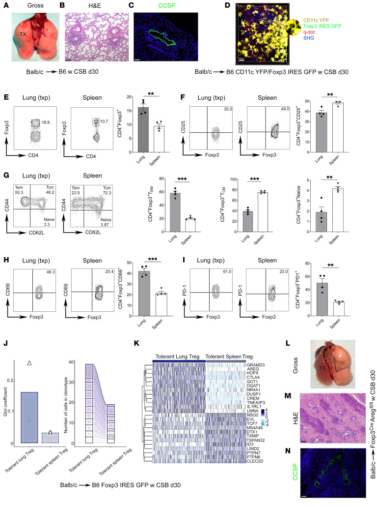
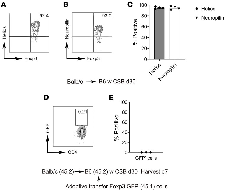
Mechanisms that mediate allograft tolerance differ between organs. We have previously shown that Foxp3+ T cell-enriched bronchus-associated lymphoid tissue (BALT) is induced in tolerant murine lung allografts and that these Foxp3+ cells suppress alloimmune responses locally and systemically. Here, we demonstrated that Foxp3+ cells that reside in tolerant lung allografts differed phenotypically and transcriptionally from those in the periphery and were clonally expanded. Using a mouse lung retransplant model, we showed that recipient Foxp3+ cells were continuously recruited to the BALT within tolerant allografts. We identified distinguishing features of graft-resident and newly recruited Foxp3+ cells and showed that graft-infiltrating Foxp3+ cells acquired transcriptional profiles resembling those of graft-resident Foxp3+ cells over time. Allografts underwent combined antibody-mediated rejection and acute cellular rejection when recruitment of recipient Foxp3+ cells was prevented. Finally, we showed that local administration of IL-33 could expand and activate allograft-resident Foxp3+ cells, providing a platform for the design of tolerogenic therapies for lung transplant recipients. Our findings establish graft-resident Foxp3+ cells as critical orchestrators of lung transplant tolerance and highlight the need to develop lung-specific immunosuppression.

Keywords: Immunology; Tolerance; Transplantation.


Figures

Figure 1
Figure 1. Allograft-resident and spleen Foxp3+ cells are phenotypically and transcriptionally distinct in tolerant lung transplant recipients.
(AC) Gross image (n = 6) (A), H&E staining (n = 6) (B), and CCSP immunofluorescence staining (n = 2) (C) of left lung transplant from BALB/c donor into CSB-treated B6 recipient, at least 30 days after engraftment. Scale bars: 100 μm. (D) Two-photon intravital microscopy of BALB/c lung at least 30 days after transplantation into CSB-treated B6 CD11c-EYFP/Foxp3-GFP recipient (n = 3). CD11c+ cells are yellow, Foxp3+ cells are green, collagen appears blue owing to second-harmonic generation (SHG), and vessels are red following i.v. injection of quantum dots. Scale bar: 20 μm. (EI) Representative flow cytometry plots and quantification of abundance of Foxp3-expressing CD45+CD90.2+CD4+CD8 T cells (E), CD25-expressing CD45+CD90.2+CD4+CD8Foxp3+ T cells (CD25 expression determined based on isotype control staining) (F), effector memory (CD44hiCD62Llo), central memory (CD44hiCD62Lhi), and naive (CD44loCD62Lhi) CD45+CD90.2+CD4+CD8Foxp3+ T cells (G), CD69-expressing CD45+CD90.2+CD4+CD8Foxp3+ T cells (H), and (I) PD-1–expressing CD45+CD90.2+CD4+CD8Foxp3+ T cells in BALB/c lung and recipient spleen at least 30 days after transplantation into CSB-treated B6 recipients (n = 4). At least 30 days after transplantation of BALB/c lungs into CSB-treated B6 Foxp3-GFP mice, CD45+CD90.2+CD4+CD8GFP+ cells were sorted and processed for TCR and genome sequencing (n = 2). (J) Gini coefficient (0 represents maximal diversity) comparison of clonal expansion (left) and number of cells in the top 11 shared clonotypes between Foxp3+ cells in tolerant lung allografts and recipient spleens (right) (triangles and circles denote individual mice). (K) Heatmap of most highly differentially expressed genes in Foxp3+ cells between tolerant lung allografts and recipient spleens (2 pooled lungs and spleens). (LN) Gross image (n = 4) (L), H&E staining (n = 4) (M), and CCSP immunofluorescence staining (n = 2) (N) of BALB/c lungs 30 days after transplantation into CSB-treated B6 Foxp3-YFP-Cre Aregfl/fl mice. Scale bars: 100 μm. Results expressed as mean ± SEM. **P < 0.01, ***P < 0.001. d30, day 30; q-dot, quantum dot; Tcm, T central memory; Tem, T effector memory; Treg, regulatory Foxp3+ T cell; TX, transplanted lung.
Figure 2
Figure 2. Graft-infiltrating Foxp3+ cells are derived from the thymus rather than from peripheral conversion of Foxp3 T cells.
(AC) Representative flow cytometric plots (A and B) and quantification (C) of Helios and neuropilin-1 expression by CD45+CD90.2+CD4+CD8Foxp3+ T cells in BALB/c lungs at least 30 days after transplantation into CSB-treated B6 recipients (n = 4). Thirty days after transplantation of BALB/c (CD45.2) lungs into CSB-treated B6 (CD45.2) recipients, CD90.2+CD4+GFP T cells, isolated from secondary lymphoid organs of B6 Foxp3-GFP (CD45.1) reporter mice, were injected i.v. Lungs were analyzed 7 days later. (D and E) Representative flow cytometric plot and analysis of GFP expression in adoptively transferred cells (CD45.1+) in lung allografts (n = 3). Results expressed as mean ± SEM.
Figure 3
Figure 3. Newly recruited Foxp3+ cells infiltrate BALT within tolerant lung allografts and differ transcriptionally from graft-resident Foxp3+ cells.
(A) BALB/c lungs initially transplanted into CSB-treated B6 CD11c-EYFP mice and then at least 30 days later retransplanted into non-immunosuppressed B6 Foxp3-GFP hosts were imaged with intravital 2-photon microscopy 3 days after retransplantation (n = 3). White arrows point to contacts between Foxp3+ (blue) and CD11c+ (green) cells within the BALT of lung allografts. Rhodamine-dextran labels vessels red. (B) BALB/c lungs initially transplanted into CSB-treated B6 Foxp3-GFP mice and then at least 30 days later retransplanted into non-immunosuppressed B6 Foxp3-RFP recipients were imaged with intravital 2-photon microscopy 3 days after retransplantation (n = 3). White arrows point to contacts between graft-resident (green) and graft-infiltrating (red) Foxp3+ cells within lung allografts. (C and D) BALB/c (CD45.2) lungs were transplanted into CSB-treated B6 (CD45.2) recipients and at least 30 days later retransplanted into non-immunosuppressed B6 (CD45.1) mice. Seven and 21 days after retransplantation, graft-resident (CD45.2) (7 days) and extravasated graft-infiltrating (CD45.1) (7 and 21 days) T cells were sorted from the lung allografts (samples were collected from 4 retransplant recipients and pooled) and processed for single-cell RNA sequencing. (C) Uniform manifold approximation and projection (UMAP) embedding plot of regulatory T cell subpopulations. (D) Heatmap of statistically significant (log2 fold change > 0.25, adjusted P value < 0.05) differentially expressed genes between graft-resident CD45.2 (day 7) and extravasated graft-infiltrating CD45.1 (days 7 and 21) regulatory T cells grouped by condition. (EI) Representative flow cytometry plots and quantification of abundance of effector memory (CD44hiCD62Llo), central memory (CD44hiCD62Lhi), and naive (CD44loCD62Lhi) graft-resident CD45.2+ versus graft-infiltrating CD45.1+CD90.2+CD4+CD8Foxp3+ T cells on day 7 (n = 4). Results expressed as mean ± SEM. Scale bars: 20 μm. **P < 0.01, ***P < 0.001. RFP, red fluorescent protein; Tcm, T central memory; Tem, T effector memory.
Figure 4
Figure 4. Depletion of recipient Foxp3+ cells results in loss of allograft tolerance.
(A) Schematic depicting left lung from BALB/c (CD45.2) donor initially transplanted into CSB-treated B6 (CD45.2) primary recipient and then at least 30 days later retransplanted into non-immunosuppressed B6 (CD45.1) or B6 Foxp3-DTR (CD45.1) secondary recipient, with diphtheria toxin (DT) administration and analysis 7 days after retransplantation. (B and C) Gross image (first panel, n = 4), H&E histology (second panel, n = 4), CCSP immunofluorescence staining (green, third panel, n = 2), and immunohistochemical staining for complement 4d (C4d) (brown, fourth panel; arrows point to alveolar endothelial C4d staining; n = 2) in DT-treated B6 (B) and B6 Foxp3-DTR (C) secondary recipients. (DF) Representative flow cytometric plots and quantification of abundance of CD69 (D), Fas (E), and IgM/IgD (F) expression in graft-infiltrating CD45.2CD45.1+CD19+B220+ B cells in control (left panels) and recipient Foxp3-depleted (right panels) retransplants (n ≥ 4). (G and H) Flow cytometric analysis of serum donor-specific IgM antibody titers (expressed as mean fluorescence intensity) (G) and International Society for Heart and Lung Transplantation (ISHLT) A rejection grades (H) in control and recipient Foxp3-depleted retransplants (n = 4). Results expressed as mean ± SEM. Scale bars: 100 μm. *P < 0.05, **P < 0.01, ***P < 0.001. DT, diphtheria toxin; DTR, diphtheria toxin receptor; TX, transplanted lung.
Figure 5
Figure 5. Rejection after recipient Foxp3+ cell depletion is dependent on B cells.
(A) BALB/c lungs were initially transplanted into CSB-treated B6 CD11c-EYFP mice and then retransplanted into non-immunosuppressed B6 Foxp3-GFP hosts at least 30 days later. Recipient-matched B6 B cells labeled with the rhodamine-based red cell dye CMTMR were injected into recipients 2 days after retransplantation, and allografts were imaged with intravital 2-photon microscopy the following day (n = 3). White arrows point to contacts between Foxp3+ (green) and B cells (red). CD11c+ cells (yellow) mark the BALT within tolerant lung allografts (n = 4). Scale bar: 20 μm. (B) Gross (top) and histological (H&E; bottom) images of BALB/c lungs that were initially transplanted into CSB-treated B6 mice and then at least 30 days later retransplanted into Foxp3-depleted non-immunosuppressed B6 recipients that received anti-CD20 (right) or isotype control antibodies (left). Scale bars: 100 μm. (C and D) Flow cytometric analysis of serum IgM DSA titers (expressed as mean fluorescence intensity) (C) and ISHLT A rejection grades (D) in retransplant recipients described in B (n ≥ 4). DT, diphtheria toxin; DTR, diphtheria toxin receptor; TX, transplanted lung.
Figure 6
Figure 6. Lung allograft tolerance is maintained in the global absence of CD4+ T cells.
(A and B) Gross (A) and histological (H&E) (B) images of BALB/c lungs that were initially transplanted into CSB-treated B6 mice and then at least 30 days later retransplanted into non-immunosuppressed B6 CD4 knockout recipients. Scale bar: 100 μm. (C and D) ISHLT A rejection grades (C) and flow cytometric analysis of serum IgM DSA titers (expressed as mean fluorescence intensity) (D) in retransplants depicted in A and B compared with tolerant BALB/c lung allografts that were retransplanted into non-immunosuppressed B6 mice (n ≥ 3). Mice were examined 7 days after retransplantation. TX, transplanted lung.
Figure 7
Figure 7. IL-33 promotes the expansion and activation of lung allograft–resident Foxp3+ cells.
BALB/c lungs were transplanted into CSB-treated B6 recipients. At least 30 days after transplantation, recipients were treated with intratracheal IL-33 or PBS, and grafts were analyzed 7 days later. (AE) Representative flow cytometry plots and quantification of abundance of Foxp3-expressing CD45+CD90.2+CD4+CD8 T cells (A), CD25-expressing CD45+CD90.2+CD4+CD8Foxp3+ T cells (B), effector memory (CD44hiCD62Llo), central memory (CD44hiCD62Lhi), and naive (CD44loCD62Lhi) CD45+CD90.2+CD4+CD8Foxp3+ T cells (C), CD69-expressing CD45+CD90.2+CD4+CD8Foxp3+ T cells (D), and (E) PD-1–expressing CD45+CD90.2+CD4+CD8Foxp3+ T cells in lung allografts after treatment with IL-33 or PBS (n = 4). (F) Gross (top) and histological (H&E; bottom) appearances of left BALB/c lungs that were initially transplanted into CSB-treated B6 recipients and then 30 days later retransplanted into DT-treated B6 Foxp3-DTR secondary recipients that received PBS (left; n = 4) or IL-33 (right; n = 9) intratracheally. Grafts were examined 7 days after retransplantation. Scale bars: 100 μm. (G and H) ISHLT A rejection grades (G) and flow cytometric analysis of serum IgM DSA titers (expressed as mean fluorescence intensity) (H) in recipients depicted in F (PBS, n = 4; IL-33, n = 9). Results expressed as mean ± SEM. *P < 0.05, **P < 0.01. DT, diphtheria toxin; DTR, diphtheria toxin receptor; Tcm, T central memory; Tem, T effector memory; TX, transplanted lung.

References

    1. Balsara KR, et al. A single-center experience of 1500 lung transplant patients. J Thorac Cardiovasc Surg. 2018;156(2):894–905. doi: 10.1016/j.jtcvs.2018.03.112. - DOI - PMC - PubMed
    1. Witt CA, et al. Lung transplant immunosuppression — time for a new approach? Expert Rev Clin Immunol. 2014;10(11):1419–1421. doi: 10.1586/1744666X.2014.959499. - DOI - PMC - PubMed
    1. Krupnick AS, et al. Central memory CD8+ T lymphocytes mediate lung allograft acceptance. J Clin Invest. 2014;124(3):1130–1143. doi: 10.1172/JCI71359. - DOI - PMC - PubMed
    1. Onyema OO, et al. Eosinophils promote inducible NOS-mediated lung allograft acceptance. JCI Insight. 2017;2(24):e96455. doi: 10.1172/jci.insight.96455. - DOI - PMC - PubMed
    1. Onyema OO, et al. Eosinophils downregulate lung alloimmunity by decreasing TCR signal transduction. JCI Insight. 2019;4(11):e128241. doi: 10.1172/jci.insight.128241. - DOI - PMC - PubMed