. 2025 Mar 18;21(3):e1012108.
doi: 10.1371/journal.ppat.1012108. eCollection 2025 Mar.

NLRP3 and AIM2 inflammasomes exacerbate the pathogenic Th17 cell response to eggs of the helminth Schistosoma mansoni

Affiliations

NLRP3 and AIM2 inflammasomes exacerbate the pathogenic Th17 cell response to eggs of the helminth Schistosoma mansoni

Madhusoodhanan Suresh Kumar Meena Kumari et al. PLoS Pathog. .

Abstract

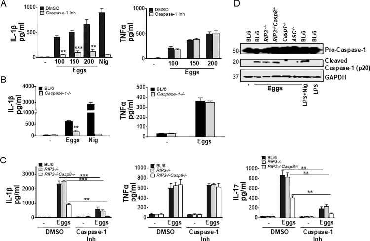
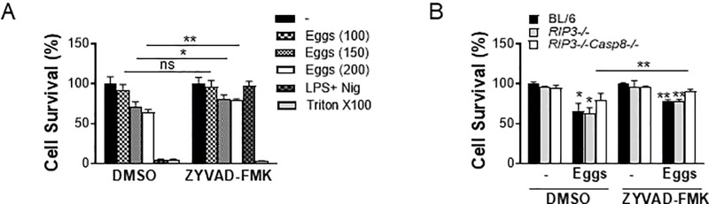
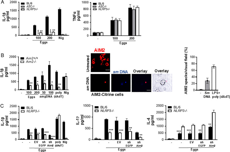
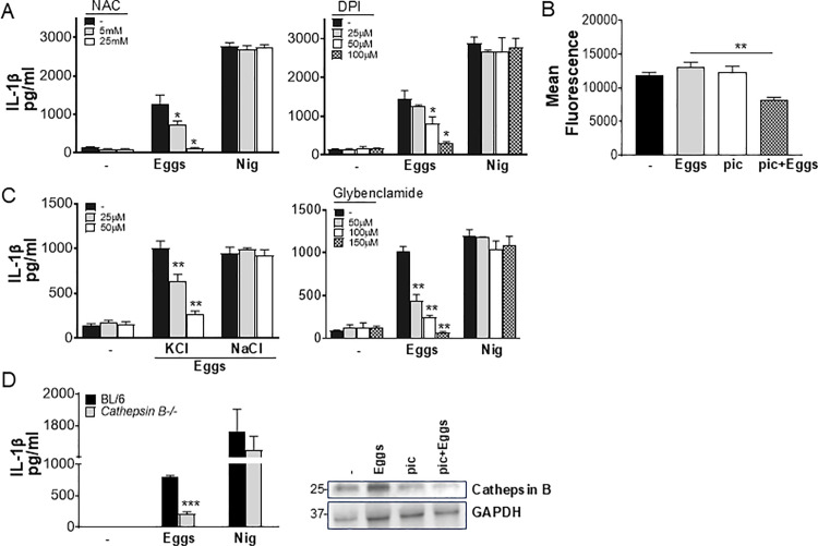
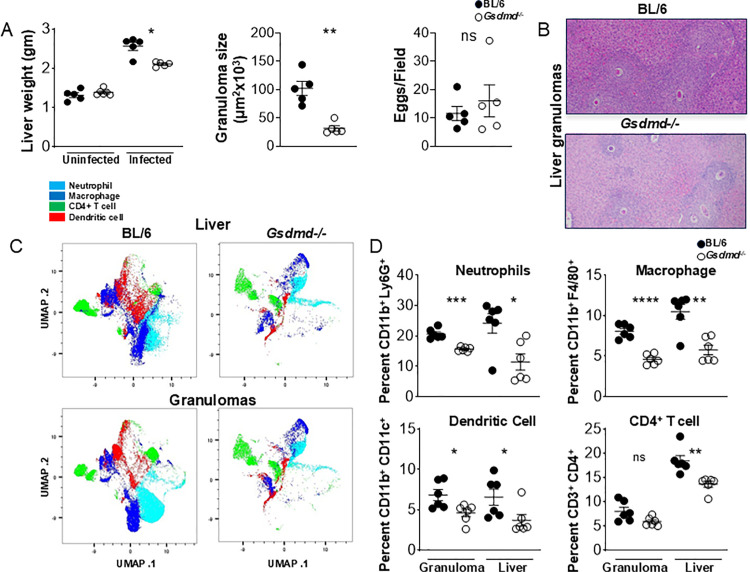
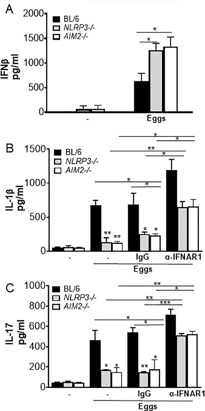
Infection with the helminth Schistosoma mansoni can cause exacerbated morbidity and mortality via a pathogenic host CD4 T cell-mediated immune response directed against parasite egg antigens, with T helper (Th) 17 cells playing a major role in the development of severe granulomatous hepatic immunopathology. The role of inflammasomes in intensifying disease has been reported; however, neither the types of caspases and inflammasomes involved, nor their impact on the Th17 response are known. Here we show that enhanced egg-induced IL-1β secretion and pyroptotic cell death required both caspase-1 and caspase-8 as well as NLRP3 and AIM2 inflammasome activation. Schistosome genomic DNA activated AIM2, whereas reactive oxygen species, potassium efflux and cathepsin B, were the major activators of NLRP3. NLRP3 and AIM2 deficiency led to a significant reduction in pathogenic Th17 responses, suggesting their crucial and non-redundant role in promoting inflammation. Additionally, we show that NLRP3- and AIM2-induced IL-1β suppressed IL-4 and protective Type I IFN (IFN-I) production, which further enhanced inflammation. IFN-I signaling also curbed inflammasome- mediated IL-1β production suggesting that these two antagonistic pathways shape the severity of disease. Lastly, Gasdermin D (Gsdmd) deficiency resulted in a marked decrease in egg-induced granulomatous inflammation. Our findings establish NLRP3/AIM2-Gsdmd axis as a central inducer of pathogenic Th17 responses which is counteracted by IFN-I pathway in schistosomiasis.


Conflict of interest statement

The authors have declared that no competing interests exist.

Figures

Fig 1
Fig 1. Egg-induced IL-1β and IL-17 production is dependent on both Caspase-1 and Caspase-8.
(A) BL/6 BMDCs were pretreated with caspase-1 inhibitor ZYVAD-FMK (50μM) for 1h before culturing for 24h with the indicated numbers of live eggs, or LPS plus nigericin (Nig). IL-1β (left) and TNFα (right) in supernatants were measured by ELISA. (B) BMDCs from BL/6 and Caspase-1-/- mice were cultured for 24h with 100 live (or no) eggs, or LPS plus Nig. IL-1β (left) and TNFα (right) in supernatants were measured by ELISA. (C) BL/6, RIP3-/- and RIP3-/-/Casp8-/- BMDCs were pretreated with ZYVAD-FMK (50μM) for 1h before culturing for 24h with 100 live (or no) eggs. IL-1β (left) and TNFα (middle) in supernatants were measured by ELISA. BL/6, RIP3-/- and RIP3-/-/Casp8-/- BMDCs were pretreated with ZYVAD-FMK (50μM) for 1h before establishing co-cultures with CD4 T cells in the presence and absence of 100 live eggs. IL-17A (right) in 72h supernatants was measured by ELISA. (D) Immunoblot analysis of pro-caspase-1 processing in whole cell lysates of BL/6, RIP3-/-, RIP3-/-/Casp8-/-, Casp1-/- and ASC-/- BMDCs left unstimulated or stimulated with 1000 eggs, LPS and Nig or LPS only. Bars represent the mean +/- SD cytokine levels of three biological replicates from one representative experiment of three with similar results. **p <0.005, ***p <0.0005.
Fig 2
Fig 2. Eggs jointly coordinate a caspase-1 and caspase-8 dependent pyroptotic cell death.
(A) BL/6 BMDCs were cultured for 24h with the indicated number of live (or no) eggs, LPS plus Nig and Triton X-100 (Tri) for 10 min. (B) BL/6, RIP3-/- and RIP3-/-/Casp8-/- BMDCs were pretreated with ZYVAD-FMK (50μM) for 1h before culturing for 24h with 100 live (or no) eggs. Cell survival was measured using calcein AM. The medium was set at 100%. Bars represent the mean +/- SD cytokine levels of three biological replicates from one representative experiment of three with similar results. *p <0.05, **p <0.005.
Fig 3
Fig 3. The loss of NLRP3 and AIM2 results in reduced Th17 and enhanced Th2 cytokine responses.
(A) BMDCs from BL/6, ASC-/- and NLRP3-/- mice were cultured for 24h with the indicated number of live (or no) eggs, or LPS plus Nig. IL-1β (left) and TNFα (right) in supernatants were measured by ELISA. (B) BMDCs from Aim2+/+ and Aim2-/- mice were cultured for 24h with the indicated number of live (or no) eggs, transfected with the indicated concentrations of Schistosoma mansoni (sm) genomic (g) DNA, LPS plus poly(dAdT), or LPS plus Nig. IL-1β in supernatants was measured by ELISA (left). Poly (dAdT) was used as an AIM2 inflammasome-activating control. (B) Confocal microscopy of LPS primed AIM2-citrine macrophages left untransfected or transfected with 100 ng/ml DAPI-labeled sm gDNA (middle). Scale bar: 20μm (top and bottom). The formation of AIM2 pyroptosomes was quantified using confocal microscopy (right). Data are representative of at least 10 fields of view and three independent experiments. (C) BL/6 and NLRP3-/- BMDCs were transduced with empty vector (EV), control shRNA against eGFP (shEGFP) and shRNA against AIM2 (shAIM2). Cells were cultured with 100 live eggs or LPS plus Nig or LPS plus poly (dAdT). IL-1β in 24h supernatants was measured by ELISA (left). The above-mentioned BMDCs were co-cultured with T cells and 100 live (or no) eggs. IL-17A (middle) and IL-4 (right) in 72h supernatants were measured by ELISA. Bars represent the mean +/- SD cytokine levels of three biological replicates from one representative experiment of three with similar results. *p <0.05, **p <0.005, ***p <0.0005.
Fig 4
Fig 4. ROS, K+ efflux and cathepsin B are required for NLRP3 activation.
(A) BL/6 BMDCs were pretreated with the indicated concentrations of ROS inhibitor N-acetyl-l-cysteine (NAC) (left) or Diphenyleneiodonium (DPI) (right) for 1h before culturing for 24h with 100 live (or no) eggs, or LPS plus Nig. IL-1β in supernatants was measured by ELISA. (B) 106 BL/6 BMDCs were pretreated with 20 μM of piceatannol (pic) for 1h before culturing for 24h with 1000 eggs. Cells were then stained with CellRox Deep Red and ROS was measured using flow cytometry (C) BL/6 BMDCs were pretreated with indicated concentrations of potassium chloride (KCl), sodium chloride (NaCl) (left) or potassium channel blocker Glybenclamide (right) for 1h before culturing for 24h with 100 live (or no) eggs, or LPS plus Nig (D) BMDCs from BL/6 and Cathepsin B-/- mice were cultured for 24h with 100 live (or no) eggs or LPS plus Nig. IL-1β in supernatants was measured by ELISA (left). Immunoblot analysis of cathepsin B in whole cell lysates of BL/6 BMDCs left unstimulated or pretreated with 20 μM piceatannol for 1h and then stimulated with 1000 eggs for 24h, stimulated with eggs, or Piceatannol only (right). Bars represent the mean +/- SD cytokine levels of three biological replicates from one representative experiment of three with similar results. *p <0.05, **p <0.005, ***p <0.0005.
Fig 5
Fig 5. Gsdmd deficiency reduces myeloid cell recruitment to the liver during schistosome infection.
(A) Weights of livers from uninfected and infected BL/6 and Gsdmd/ mice (left). Granuloma size was determined by morphometric analysis. Each dot represents average granuloma size of 10–20 granulomas in two sections from individual mice (middle). Number of eggs per 2.4 mm2 field assessed on H&E-stained liver sections at 100× magnification. An average of 20 fields per liver section was assessed. Images are representative of three independent experiments (right). (B) Representative histopathology of liver granulomas of BL/6 and Gsdmd/ mice (magnification, 100×). (C) UMAP of neutrophils, macrophages, CD4+T cells, and dendritic cells isolated from livers of C57BL/6 and Gsdmd-/- mice infected with Schistosoma mansoni for 7 weeks. (D) Percentages of liver- and granuloma-derived neutrophils, macrophages, dendritic cells and CD4+ T cells as a proportion of CD45+ cells: (top left) Ly6G+ cells, (top right) F4/80+ cells, (bottom left) CD11b+ CD11c+ cells, (bottom right) CD3+CD4+ cells. Data are representative of two independent experiments. Significance was determined using Student’s t-test *p < 0.05, **p < 0.005, ***p < 0.0005, ****p < 0.00005; ns, not significant.
Fig 6
Fig 6. Blocking IFNAR leads to enhancement of IL-1β and IL-17 production.
(A) BMDCs from BL/6, NLRP3-/- and AIM2-/- BMDCs were cultured for 24h with 100 live (or no) eggs. IFNβ in supernatants was measured by ELISA. (B) BL/6, NLRP3-/- and AIM2-/- BMDCs were pretreated with IFN-I receptor blocking antibody (α-IFNAR1) (20μg/ml) or control IgG (20μg/ml) for 1h before culturing for 24h with 100 live (or no) eggs. IL-1β in supernatants was measured by ELISA. (C) The above mentioned BMDCs were co-cultured with T cells together with 100 live (or no) eggs. IL-17A in 72h supernatants were measured by ELISA. Bars represent the mean +/- SD cytokine levels of three biological replicates from one representative experiment of three with similar results. *p <0.05, **p <0.005, ***p <0.0005.

Update of

Similar articles

References

    1. Lo NC, Bezerra FSM, Colley DG, Fleming FM, Homeida M, Kabatereine N, et al.. Review of 2022 WHO guidelines on the control and elimination of schistosomiasis. Lancet Infect Dis. 2022;22(11):e327-35. - PubMed
    1. Hernandez HJ, Wang Y, Tzellas N, Stadecker MJ. Expression of class II, but not class I, major histocompatibility complex molecules is required for granuloma formation in infection with Schistosoma mansoni. Eur J Immunol. 1997;27(5):1170–6. doi: 10.1002/eji.1830270518 - DOI - PubMed
    1. Iacomini J, Ricklan DE, Stadecker MJ. T cells expressing the gamma delta T cell receptor are not required for egg granuloma formation in schistosomiasis. Eur J Immunol. 1995;25(4):884–8. doi: 10.1002/eji.1830250404 - DOI - PubMed
    1. Phillips SM, DiConza JJ, Gold JA, Reid WA. Schistosomiasis in the congenitally athymic (nude) mouse. I. Thymic dependency of eosinophilia, granuloma formation, and host morbidity. J Immunol. 1977;118(2):594–9. doi: 10.4049/jimmunol.118.2.594 - DOI - PubMed
    1. Molehin AJ, McManus DP, You H. Vaccines for Human Schistosomiasis: Recent Progress, New Developments and Future Prospects. Int J Mol Sci. 2022;23(4). - PMC - PubMed

MeSH terms

LinkOut - more resources