

Gamma knife radiosurgery for a rare Rosette-forming glioneuronal tumor in the brainstem region: A case report and literature review
- PMID: 40101077
- PMCID: PMC11922452
- DOI: 10.1097/MD.0000000000041869
Gamma knife radiosurgery for a rare Rosette-forming glioneuronal tumor in the brainstem region: A case report and literature review
Abstract
Rationale: Rosette-forming glioneuronal tumor (RGNT) is a rare primary nervous system tumor, with limited treatment guidelines due to its rarity, especially in the brainstem. This report presents a unique case of brainstem RGNT treated with gamma knife radiosurgery (GKRS).
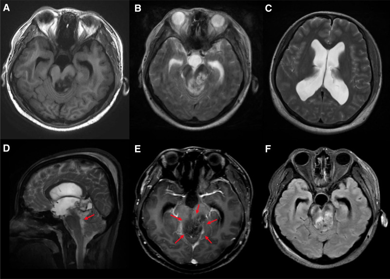
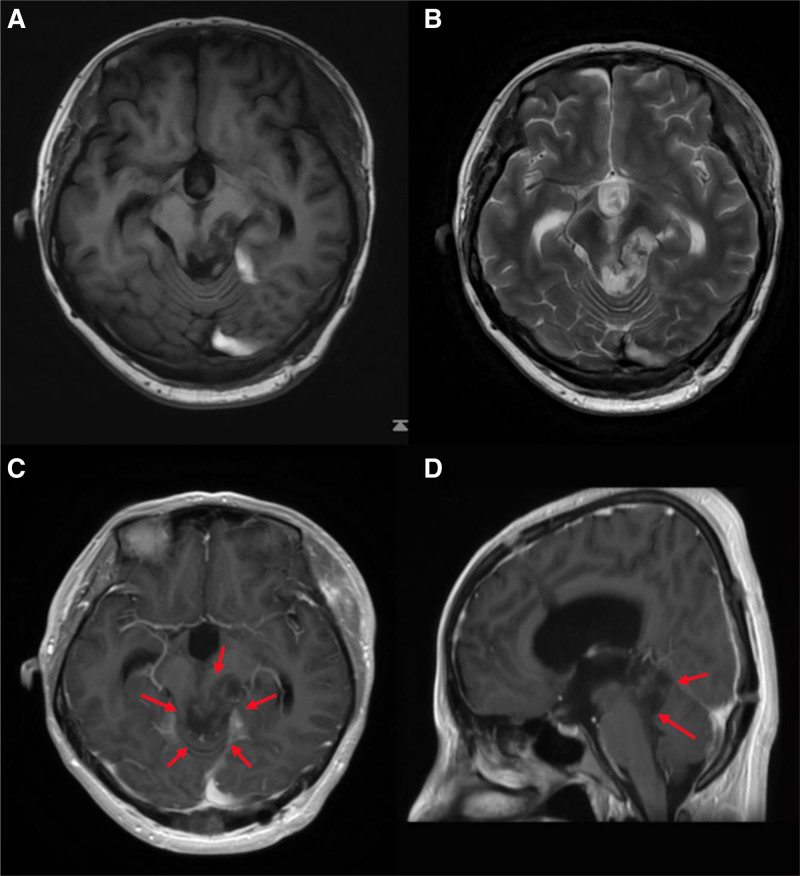
Patient concerns: A 35-year-old woman sought medical attention after sudden syncope and rapid decline in consciousness. Magnetic resonance imaging revealed a mass in the pineal region, extending to the brainstem and thalamus. Due to the critical location, only partial resection of the pineal tumor was possible, leaving most of the residual tumor in the vital brainstem area, requiring urgent intervention to control its growth and prevent sudden complications.
Diagnoses: Postoperative histopathological results confirmed a diagnosis of RGNT.
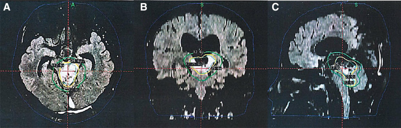
Interventions: The patient underwent 25 Gy/5 fractions of GKRS using the frameless Gamma Knife ICON™ (Elekta) device, as confirmed by cone-beam computed tomography scans for precise dose distribution and patient alignment.
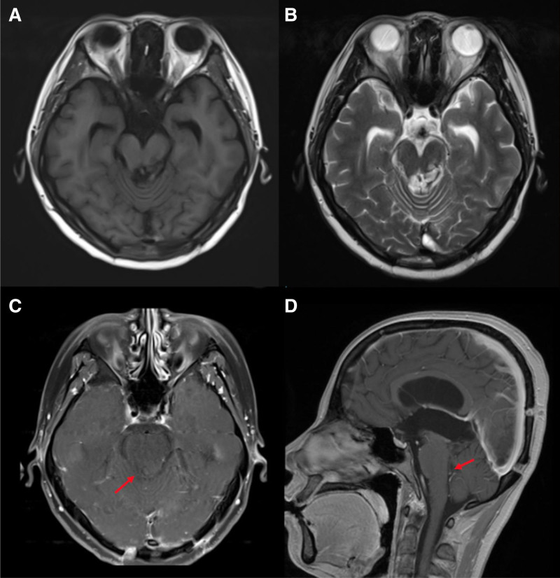
Outcomes: GKRS was performed successfully and safely. The tumor significantly shrank 3 months post-GKRS, and the patient experienced symptom relief without any adverse effects.
Lessons: GKRS is considered an effective modality for RGNT in high-risk brainstem areas, minimizing risks while controlling tumor growth and alleviating symptoms. In addition, the frameless Gamma Knife ICON™ device enhanced patient comfort and treatment precision. GKRS offers a noninvasive alternative for similar RGNT cases.
Copyright © 2025 the Author(s). Published by Wolters Kluwer Health, Inc.
Conflict of interest statement
The authors have no funding and conflicts of interest to disclose.
Figures
Similar articles
-
Staged Gamma Knife radiosurgery for a rosette-forming glioneuronal tumor of the fourth ventricle: a case report.Childs Nerv Syst. 2023 Nov;39(11):3323-3326. doi: 10.1007/s00381-023-06014-y. Epub 2023 Jun 5. Childs Nerv Syst. 2023. PMID: 37272935
-
Gamma knife radiosurgery for symptomatic brainstem intra-axial cavernous malformations.World Neurosurg. 2013 Dec;80(6):e261-6. doi: 10.1016/j.wneu.2012.09.013. Epub 2012 Sep 22. World Neurosurg. 2013. PMID: 23010066
-
Fractionated Gamma Knife radiosurgery for skull base meningiomas: a single-institution experience.Neurosurg Focus. 2019 Jun 1;46(6):E8. doi: 10.3171/2019.3.FOCUS1963. Neurosurg Focus. 2019. PMID: 31153152
-
Single-Fraction Gamma-Knife Radiosurgery with or without Previous Surgery for Cavernous Sinus Meningiomas: A Single-Center Experience and Systematic Literature Review.Niger J Clin Pract. 2023 May;26(5):545-551. doi: 10.4103/njcp.njcp_2033_21. Niger J Clin Pract. 2023. PMID: 37357468
-
Significant Hemorrhage Rate Reduction after Gamma Knife Radiosurgery in Symptomatic Cavernous Malformations: Long-Term Outcome in 95 Case Series and Literature Review.Stereotact Funct Neurosurg. 2017;95(6):369-378. doi: 10.1159/000480664. Epub 2017 Nov 4. Stereotact Funct Neurosurg. 2017. PMID: 29131117 Review.
References
-
- Komori T, Scheithauer BW, Hirose T. A rosette-forming glioneuronal tumor of the fourth ventricle: infratentorial form of dysembryoplastic neuroepithelial tumor? Am J Surg Pathol. 2002;26:582–91. - PubMed
-
- Schlamann A, Von Bueren AO, Hagel C, et al. . An individual patient data meta-analysis on characteristics and outcome of patients with papillary glioneuronal tumor, rosette glioneuronal tumor with neuropil-like Islands and rosette forming glioneuronal tumor of the fourth ventricle. PLoS One. 2014;9:e101211. - PMC - PubMed
Publication types
MeSH terms
LinkOut - more resources
Full Text Sources
