. 2025 Mar 18;13(3):e010179.
doi: 10.1136/jitc-2024-010179.

Progesterone receptor-dependent downregulation of MHC class I promotes tumor immune evasion and growth in breast cancer

Affiliations

Progesterone receptor-dependent downregulation of MHC class I promotes tumor immune evasion and growth in breast cancer

Julio C Tinoco et al. J Immunother Cancer. .

Abstract

Background: Breast cancer (BC) continues to be a major health concern with 250,000 new cases diagnosed annually in the USA, 75% of which are hormone receptor positive (HR+), expressing estrogen receptor alpha (ER) and/or the progesterone receptor (PR). Although ER-targeted therapies are available, 30% of patients will develop resistance, underscoring the need for new non-ER/estrogen-based treatments. Notably, HR+BCs exhibit poor lymphocyte infiltration and contain an immunosuppressive microenvironment, which contributes to the limited efficacy of immunotherapies in HR+BC. In this study, we demonstrate that PR/progesterone signaling reduces major histocompatibility complex (MHC) Class I expression, facilitating immune evasion and escape from immune-based clearance of PR+tumors.

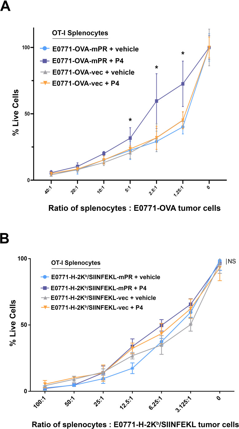
Methods: To determine the effect of PR/progesterone on MHC Class I expression, we treated human and mouse mammary tumor cell lines with progesterone and/or interferon (IFN) and measured expression of genes involved in antigen processing and presentation (APP), as well as surface MHC Class I expression. We used the OT-I/SIINFEKL model antigen system to measure the impact of progesterone on immune cell-mediated killing of modified tumor cells. We also analyzed two large BC clinical cohorts to determine how PR expression correlates with APP gene expression and MHC Class I expression in ER-positive tumors.

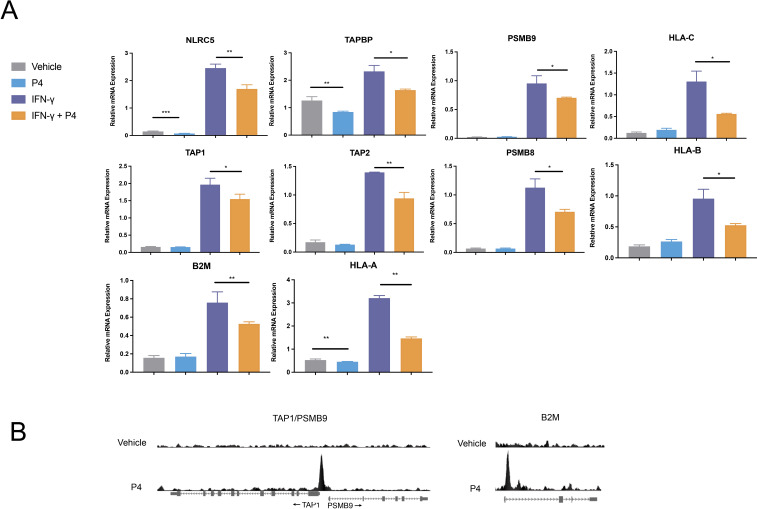
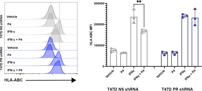
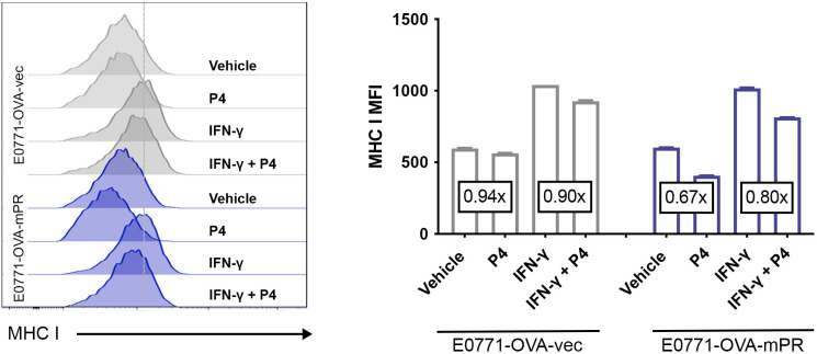
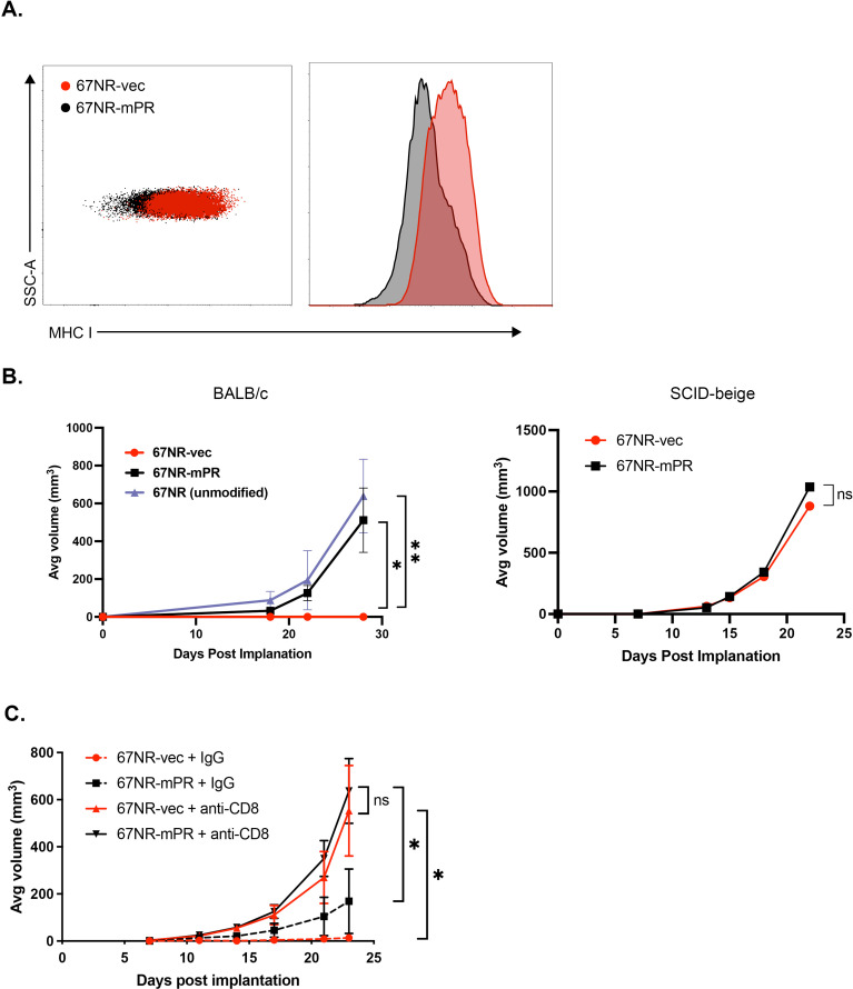
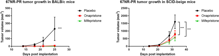
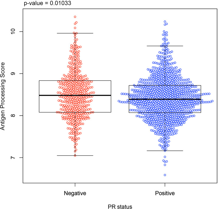
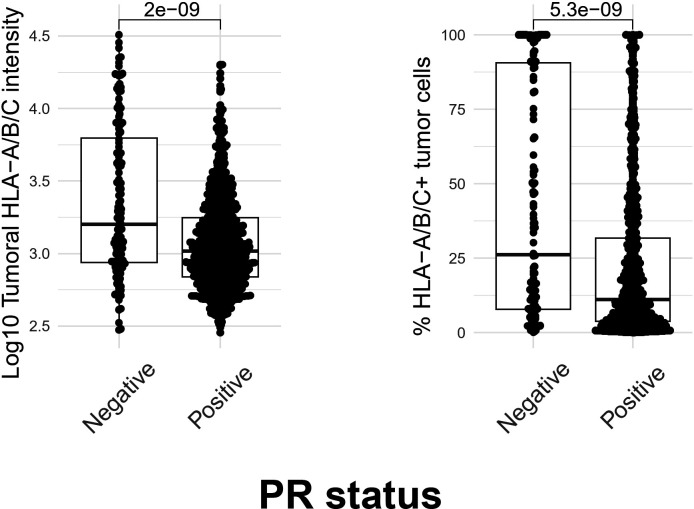
Results: In vitro, we show that PR/progesterone signaling reduces APP gene expression and MHC class I expression in human and breast mammary tumor cell lines. PR-mediated attenuation of APP/MHC Class I expression is more pronounced in the presence of IFN. In immune cell killing assays, PR-expressing mammary tumor cells treated with progesterone are protected from immune-mediated cytotoxicity. We demonstrate that PR expression in vivo prevents immune-mediated rejection of xenoantigen-modified mammary tumor cell lines through mechanisms involving MHC Class I expression and CD8 T cells. Data analysis of two large BC cohorts reveals lower APP gene expression and MHC Class I expression in ER/PR-positive tumors compared with ER-positive/PR-negative tumors. These findings show that HR+BCs, specifically PR+tumors, downregulate APP/MHC class I machinery through PR/progesterone signaling. Use of pharmacological PR/progesterone inhibitors may reverse these effects in patients with BC, thereby improving immunosurveillance and response to immunotherapies.

Keywords: Breast Cancer; Immunosuppression; Major histocompatibility complex - MHC.


Conflict of interest statement

Competing interests: CRH received funds from Context Therapeutics (former developer of onapristone). JMB receives research support from Genentech/Roche and Incyte Corporation, has received advisory board payments from AstraZeneca, Eli Lilly, and Mallinckrodt, and is an inventor on patents regarding immunotherapy targets and biomarkers in cancer. The remaining authors have nothing to disclose.

Figures

Figure 1
Figure 1. PR/progesterone signaling represses interferon response and attenuates APP/MHC class I machinery in human breast cancer cells. (A) T47D ER/PR positive cells were pretreated with progesterone (P4, or vehicle control) at a concentration of 100 nM for 6 hours, then treated with recombinant human interferon gamma (IFN-γ; or vehicle control) at 1,000 U/mL for 12 hours. Following treatment, the cells were harvested, and the gene expression of APP/MHC class I was measured by quantitative PCR (n=3) and normalized to an internal control (B-actin). Error bars represent the SEM between replicates. Statistical differences among the groups were assessed using 2-way ANOVA followed by Tukey’s post hoc tests for pairwise comparisons; relevant comparisons are highlighted. **p≤0.01, ***p≤0.001, **p≤0.01, *p≤0.05. (B) Progesterone-induced PR binding sites in T47D cells shown for APP genes where PR binding was detected within 10 kb of the start site of the gene. ANOVA, analysis of variance; APP, antigen processing and presentation; ER, estrogen receptor; MHC, major histocompatibility complex; mRNA, messenger RNA; PR, progesterone receptor.
Figure 2
Figure 2. PR-dependent downregulation of MHC class I expression in breast cancer cells. T47D NS (non-silencing) and PR shRNA cells were treated with progesterone (P4) at a concentration of 100 nM, recombinant human interferon gamma (IFN-γ) at 10 U/mL, or a combination of P4 and IFN-γ for 48 hours. Following treatment, the cells were harvested, and the expression of HLA-ABC was assessed using flow cytometry (histograms on left, quantified on right). Error bars represent the SD between triplicate replicates. Statistical differences among groups were assessed using 2-way ANOVA followed by Tukey’s post hoc tests for pairwise comparisons and relevant differences are highlighted on the graph; **p≤0.01. ANOVA, analysis of variance; MFI, mean fluorescence intensity; MHC, major histocompatibility complex; PR, progesterone receptor; shRNA, short hairpin RNA.
Figure 3
Figure 3. PR/progesterone signaling attenuates MHC class I in mouse mammary tumor cells expressing PR. E0771-OVA-vec and E0771-OVA-mPR cells were treated with progesterone (20 nM; P4) or recombinant mouse interferon gamma (IFN-γ; 100 ng/mL). MHC class I expression was assessed using flow cytometry 48 hours post-treatment (histograms on left, quantified on right). Error bars represent the SD between replicates. Fold change between relevant treatment groups is shown in text boxes. Statistical differences among groups were assessed using 2-way ANOVA followed by Tukey’s post hoc tests for pairwise comparisons. All pairwise comparisons are significant (p≤0.05 or less), with the exception of vehicle treatment between cell lines. ANOVA, analysis of variance; MFI, mean fluorescence intensity; MHC, major histocompatibility complex; PR, progesterone receptor.
Figure 4
Figure 4. PR expression in tumor cells shields from immune-mediated killing. (A) Splenocytes were isolated from OT-I C57BL/6 mice and cultured with E0771-OVA-vec or E0771-mPR for 3 days. The media were supplemented with 20 nM progesterone or control. After 72 hours, non-adherent cells were removed, remaining live cells were lysed, and luciferase activity was measured. All conditions included n=6 replicates, with error bars representing the SD. Statistical significance between groups at each ratio was determined using multiple t-tests with Bonferroni correction; *p≤0.05. (B) Assay performed as in (A), with E0771-H-2Kb expressing E0771 variants. Statistical significance between groups at each ratio was determined using multiple t-tests with Bonferroni correction. All comparisons were NS=not significant, with the exception of two individual data points (these data points are marked with a star as the data point marker, instead of a circle, square, or triangle); *p≤0.05. PR, progesterone receptor.
Figure 5
Figure 5. PR-induced MHC I downregulation facilitates immune evasion from CD8+T cells. (A) Flow cytometric analysis of MHC I expression on untreated 67NR tumor cells stably expressing either empty vector (67NR-vec) or mouse PR (67NR-mPR). (B) 67NR-vec, 67NR-mPR, or unmodified 67NR parental cells were injected into the mammary fat pads of immunocompetent BALB/c mice (left panel) or immunocompromised SCID-beige mice (right panel). (C) 67NR-vec or 67NR-mPR cells were injected into BALB/c mice and treated with either anti-CD8 monoclonal antibodies (mAb) or IgG control biweekly for 4 weeks. Error bars represent STD. Differences in tumor growth over time among the groups were analyzed using linear mixed models for repeated measure data using SAS procedure GLIMMIX AR(1) covariance structure (see Methods); *p≤0.05, **p≤0.01. MHC, major histocompatibility complex; ns, not significant; PR, progesterone receptor; STD, standard deviation.
Figure 6
Figure 6. Inhibition of PR+tumor growth by antiprogestins is immune-mediated. 30 mg mifepristone, onapristone, or placebo pellets were implanted subcutaneously into the neck of immunocompetent BALB/c mice (left panel) or immunocompromised SCID-beige mice (right panel). 7 days following pellet insertion, 67NR-mPR cells were injected into the mammary fat pads of recipient mice, and tumor growth was measured over time. Error bars represent STD. Differences in tumor growth over time among the groups were analyzed using linear mixed models for repeated measure data using SAS procedure GLIMMIX AR(1) covariance structure (see Methods); ***p≤0.001. PR, progesterone receptor; STD, standard deviation.
Figure 7
Figure 7. APP gene expression is lower in PR-positive human breast tumors. The antigen processing and presentation (APP) gene score (see Methods) is shown for all ER-positive tumors in the METABRIC database, sorted by PR positivity; PR-negative (red) and PR-positive (blue). APP genes included in the composite gene score: HLA-A, HLA-B, HLA-C, NLRC5, B2M, TAP1, TAP2, TAPBP, PSMB8, and PSMB9). The statistical test was carried out using a Wilcoxon rank-sum test. ER, estrogen receptor; METABRIC, Molecular Taxonomy of Breast Cancer International Consortium; PR, progesterone receptor.
Figure 8
Figure 8. MHC Class I expression is lower in PR-positive human breast tumors. Tumor-specific MHC-I (HLA-A/B/C) expression was measured using immunofluorescent assays staining for CK and MHC-I (HLA-A/B/C) on formalin-fixed paraffin-embedded tissue sections from patients in the CBCS cohort. To evaluate the association between PR and MHC-I expression, we performed a two-sample t-test comparing log10 normalized MHC-I fluorescent intensity (left) and the percentage of MHC-I positive tumor cells (right) between PR positive and negative tumors (all ER positive), representing 992 tumors. P values are reported above each comparison. CBCS, The Carolina Breast Cancer Study; ER, estrogen receptor; MHC, major histocompatibility complex; PR, progesterone receptor.

Similar articles

Cited by

References

    1. Atlanta: American Cancer Society; 2024. Cancer facts & figures 2024.
    1. Li Y, Yang D, Yin X, et al. Clinicopathological Characteristics and Breast Cancer-Specific Survival of Patients With Single Hormone Receptor-Positive Breast Cancer. JAMA Netw Open. 2020;3:e1918160. doi: 10.1001/jamanetworkopen.2019.18160. - DOI - PMC - PubMed
    1. A Comparison of Letrozole and Tamoxifen in Postmenopausal Women with Early Breast Cancer. N Engl J Med. 2005;353:2747–57. doi: 10.1056/NEJMoa052258. - DOI - PubMed
    1. Hanker AB, Sudhan DR, Arteaga CL. Overcoming Endocrine Resistance in Breast Cancer. Cancer Cell. 2020;37:496–513. doi: 10.1016/j.ccell.2020.03.009. - DOI - PMC - PubMed
    1. Beral V, Study CMW. Breast cancer and hormone-replacement therapy in the Million Women Study. The Lancet. 2003;362:419–27. doi: 10.1016/S0140-6736(03)14065-2. - DOI - PubMed

Substances