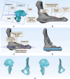
Anatomical Study of Lateral Compression II Screw Path and Entry Parameters Based on Three-Dimensional CT Image Reconstruction Techniques
- PMID: 40102032
- PMCID: PMC12050174
- DOI: 10.1111/os.70011
Anatomical Study of Lateral Compression II Screw Path and Entry Parameters Based on Three-Dimensional CT Image Reconstruction Techniques
Abstract
Objective: Lateral compression II (LC-II) fractures, a common type of pelvic injury, often require closed reduction and percutaneous screw fixation due to posterior pelvic ring instability. However, existing methods fail to adequately account for the internal structure of the screw path and lack precise anatomical guidance, increasing surgical risks. This study utilized digital medical software to analyze the LC-II screw path and entry parameters, providing the anatomical references.
Methods: This retrospective study enrolled 43 adult patients (21 males and 22 females) who underwent a complete computed tomography (CT) scan examination from February 2017 to February 2019. The digital three-dimensional (3D) pelvic model was reconstructed, and the ideal LC-II screw path was designed by the cross-section method. The primary evaluation parameters included the screw path length (D AP), maximum diameter (D max), distances at narrow points (D1 and D2), bone thickness parameters (OW1 and IW1; OW2 and IW2), and screw entry angles (∠α, ∠β, ∠γ).
Results: Of 43 patients, 42 successfully completed LC-II screw path construction. Among 21 female patients, 5 (23.8%) could accommodate screws with a maximum diameter of < 6.5 mm. Compared with female patients, male patients exhibited significantly higher D AP, D max, D 2, OW1, IW1, IW1/OW1, and IW2/OW2 (p < 0.05). The ∠γ was significantly lower in male patients. Furthermore, digital 3D pelvic model observations revealed that LC-II screws bone entry points in the anterior iliac region were all located posterior to the anterior inferior iliac spine (AIIS). The angles between the LC-II screw and coronal plane were 48.06° in males and 45.10° in females, while the angles between the LC-II screw and sagittal plane were 27.14° and 25.60°, respectively.
Conclusion: This study utilized digital medical software to construct the LC-II screw path and analyze sex-based differences, highlighting the importance of individualized preoperative path planning and providing essential anatomical evidence for the precise and safe percutaneous insertion of LC-II screws.
Keywords: anatomy; fracture fixation; lateral compression II screw; mimics software; pelvic fracture.
© 2025 The Author(s). Orthopaedic Surgery published by Tianjin Hospital and John Wiley & Sons Australia, Ltd.
Conflict of interest statement
The authors declare no conflicts of interest.
Figures


Similar articles
-
A new technique for percutaneous screw fixation for treating FFP IIIa and IIIb fragility fractures of the pelvis.Sci Rep. 2024 Jul 30;14(1):17681. doi: 10.1038/s41598-024-68201-4. Sci Rep. 2024. PMID: 39085304 Free PMC article.
-
Calculation of CT ideal screw path and safety angle before percutaneous sacroiliac screw placement.Arch Orthop Trauma Surg. 2025 Feb 1;145(1):153. doi: 10.1007/s00402-025-05774-3. Arch Orthop Trauma Surg. 2025. PMID: 39891735 Free PMC article.
-
[Implantation of iliosacral screws. Simulation of optimal placement by 3-dimensional X-ray computed tomography].Rev Chir Orthop Reparatrice Appar Mot. 2000 Jun;86(4):360-9. Rev Chir Orthop Reparatrice Appar Mot. 2000. PMID: 10880935 French.
-
2D versus 3D fluoroscopy-based navigation in posterior pelvic fixation: review of the literature on current technology.Int J Comput Assist Radiol Surg. 2017 Jan;12(1):69-76. doi: 10.1007/s11548-016-1465-5. Epub 2016 Aug 8. Int J Comput Assist Radiol Surg. 2017. PMID: 27503119 Review.
-
Percutaneous sacroiliac screw fixation with a 3D robot-assisted image-guided navigation system : Technical solutions.Oper Orthop Traumatol. 2025 Feb;37(1):3-13. doi: 10.1007/s00064-024-00871-9. Epub 2024 Nov 18. Oper Orthop Traumatol. 2025. PMID: 39556213 Free PMC article. Review.
References
-
- Starr A. J., Walter J. C., Harris R. W., Reinert C. M., and Jones A. L., “Percutaneous Screw Fixation of Fractures of the Iliac Wing and Fracture‐Dislocations of the Sacro‐Iliac Joint (OTA Types 61‐B2.2 and 61‐B2.3, or Young‐Burgess ‘Lateral Compression Type II’ Pelvic Fractures),” Journal of Orthopaedic Trauma 16, no. 2 (2002): 116–123. - PubMed
-
- Berry J. L., Stahurski T., and Asher M. A., “Morphometry of the Supra Sciatic Notch Intrailiac Implant Anchor Passage,” Spine (Phila Pa 1976) 26, no. 7 (2001): E143–E148. - PubMed
-
- Yang G. Q. R. M., Du X. Y., Yin X. J., and Zhang Y. Q., “Anatomic Study of Corona Mortis Artery by CT Angiography Combined With Three‐Dimensional Reconstruction Technology,” Chinese Journal of Anatomy and Clinics 26, no. 1 (2021): 45–49.
-
- Xu Y. J., He X. Q., Xu Y. Q., et al., “Anatomical Study of Popliteal Artery Branch Variation Based on CT Angiography of the Lower Extremity,” Chinese Journal of Anatomy and Clinics 25, no. 5 (2020): 472–477.
-
- Zhang Y. Z., Liu G., Zhang L. F., Mo W. P., Xu Z. G., and J Y. F., “Digital Analysis and Certification of Osseous Pathway for Anterograde Screwing in Acetabular Posterior Column,” Chinese Journal of Orthopaedic Trauma 20, no. 5 (2018): 389–393.
MeSH terms
Grants and funding
LinkOut - more resources
Full Text Sources
Medical

