. 2025 May;37(5):e70019.
doi: 10.1111/jne.70019. Epub 2025 Mar 18.

Comprehensive chemoanatomical mapping, and the gonadal regulation, of seven kisspeptin neuronal populations in the mouse brain

Affiliations

Comprehensive chemoanatomical mapping, and the gonadal regulation, of seven kisspeptin neuronal populations in the mouse brain

Vito S Hernández et al. J Neuroendocrinol. 2025 May.

Abstract

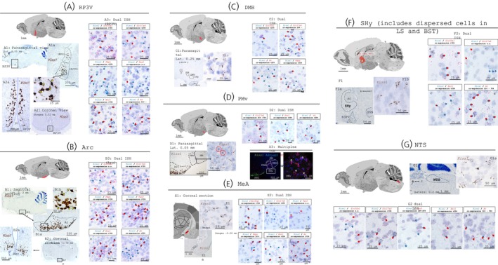
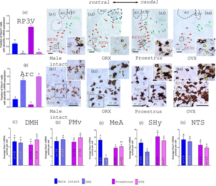
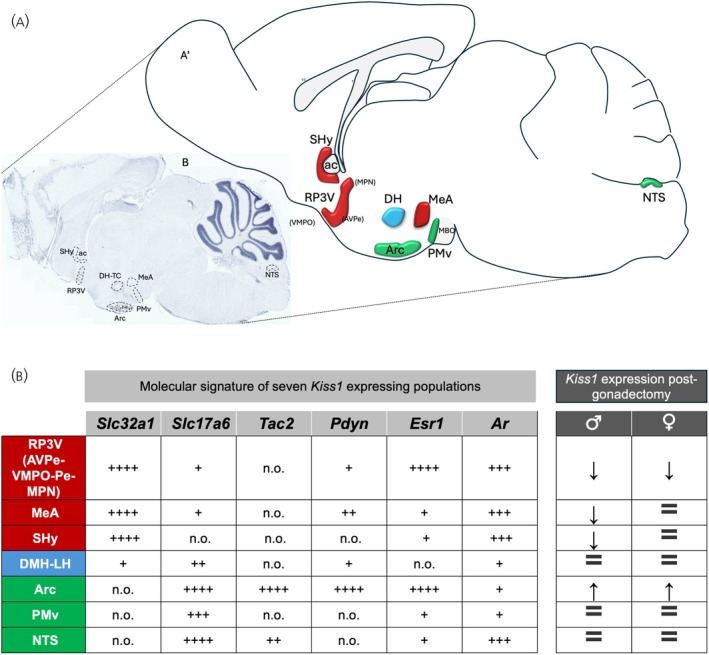
Kisspeptinergic signaling is well-established as crucial for the regulation of reproduction, but its potential broader role in brain function is less understood. This study investigates the distribution and chemotyping of kisspeptin-expressing neurons within the mouse brain. RNAscope single, dual, and multiplex in situ hybridization methods were used to assess kisspeptin mRNA (Kiss1) expression and its co-expression with other neuropeptides, excitatory and inhibitory neurotransmitter markers, and sex steroid receptors in wild-type intact and gonadectomized young adult mice. Seven distinct kisspeptin neuronal chemotypes were characterized, including two novel kisspeptin-expressing groups described for the first time, that is, the Kiss1 population in the ventral premammillary nucleus and the nucleus of the solitary tract. Kiss1 mRNA was also observed to localize in both somatic and dendritic compartments of hypothalamic neurons. High androgen receptor expression and changes in medial amygdala and septo-hypothalamic Kiss1 expression following GDX in males, but not in females, suggest a role for androgen receptors in regulating kisspeptin signaling. This study provides a detailed chemoanatomical map of kisspeptin-expressing neurons, highlighting their potential functional diversity. The discovery of a new kisspeptin-expressing group and gonadectomy-induced changes in Kiss1 expression patterns suggest broader roles for kisspeptin in brain functions beyond those of reproduction.

Keywords: Ar; Esr1; gonadectomy; neuropeptides; vesicular amino‐acid transporter.


Conflict of interest statement

The authors declare no conflicts of interest.

Figures

FIGURE 1
FIGURE 1
Anatomical localization and chemotyping of seven Kiss1‐expressing neuronal populations in the intact mouse brain (A and B: From female mice in proestrus and C‐G from male mice). (A) KP population in the rostro‐periventricular area of the hypothalamus (KPRP3V) shown in a parasagital (A1 and inset) and in a coronal view (A2) have a heterogeneous spatial distribution in the rostro‐caudal axis and a steep decrease in density in the medial to lateral direction. The yellow arrows in the figures indicate cells with prominent expression of Kiss1 mRNA in the dendritic compartment. A3 panels show RNAscope duplex reactions that demonstrate the expression of Kiss1 (blue arrows), and other mRNAs (red arrows) coding for Slc32a1, Slc17a6, Tac2, Pdyn, Esr1 or Ar. Double arrows show that in this region Kiss1 is co‐expressed with Slc32a1, Pdyn, Esr1 and Ar. The average ratio of co‐expression for anatomical structure per probe combination is expressed in quartile intervals for each micrograph. (B) Kisspeptin population in the arcuate nucleus of the hypothalamus (KPARC). B1b and inset show Kiss1 expressing neurons with dendritic Kiss1 expression (yellow arrows). B3 parasagittal micrographs show that in this population Kiss1 is colocalized with Slc17a6, Tac2, Pdyn, Esr1, and Ar mRNAs but not with Slc32a1. (C) Kiss1 population in the dorsomedial hypothalamic area (KPDMH). Kiss1 detected by singleplex RNAscope is shown in a parasagittal section in C1, Kiss1‐expressing neurons in the DMH are indicated by brown arrows in the inset. Duplex RNAscope micrographs in C2 show that in this population Kiss1 is colocalized with Slc17a6, Slc32a1, Pdyn, and AR. No co‐expression with Tac2, or Esr1 mRNAs was observed. (D) Kisspeptin mRNA expressing neuronal population in the ventral premammillary nucleus (KPPMv). D1 and inset: Micrograph of the PMv, taken from a parasagittal section shows Kiss1 expressing neurons scattered in the PMv, the expression levels of Kiss1 were low. DISH (D2) and MISH (D3) RNAscope reactions show that this newly characterized population of Kiss1 neurons co‐express the mRNAs for Slc17a6 (D2a), Esr1 (D2b), AR (D2c), PACAP and neurotensin (Adcyap1 and Nts respectively, D3). (E) Kiss1 expressing population in the medial amygdala (KPMeA). Panel E1, low and high magnification micrographs of a coronal section showing Kiss1 expressing neurons concentrated in the posterodorsal part of the medial amygdala (MeApd). E2, Duplex RNAscope showed that Kiss1 neurons strongly co‐expressed Slc32a1(E2a) and Ar (E2f) and to a lower level also expressed Slc17a6 (E2b), Esr1 (E2e), and Pdyn (E2d). (F) Kiss1 expressing population in the septo‐hypothalamic area (KPSHy). Panel F1, parasagittal section depicts the SHy area, spanning septal, BNST, and hypothalamic regions where scattered low Kiss1‐expressing neurons (inset of F1) were found around the anterior commissure. F2, micrographs taken from SHy in slices treated for RNAscope Duplex. Kiss1 was observed to be colocalized with Slc32a1 (F2a), Esr1 (F2c), and Ar (F2d). No co‐expression with Tac2, Pdyn, or Slc17a6 (F2b) mRNAs was observed in this region. (G) Kiss1 expressing population in the NTS (KPNTS). Singleplex RNAscope (G1), showed Kiss1 expressing neurons located in the solitary tract nucleus. G1 and G1a, photomicrograph of a sagittal section depicting neurons expressing Kiss1 at low abundance. G2 panels show by RNAscope Duplex the co‐expression of Kiss1 mRNA with Slc17a6 (G2b), Tac2 (G2c), Esr1 (G2d), and Ar (G2e), no co‐expression with Slc32a1 (G2a) or Pdyn was observed.
FIGURE 2
FIGURE 2
Effect of gonadectomy on Kiss1 expression in the seven Kiss1 expressing populations of the mouse brain. (A, B) The effects of gonadectomy were quantified in the two major population of Kiss1 expressing neurons, namely the RP3V (panels As) and Arc (panels Bs). The neurons in these regions showed a high cellular expression of Kiss1, and an heterogeneous spatial distribution. These two populations were distinguished by the expression of Kiss1 in the dendritic compartment. Thus we measured the fraction of Kiss1 cells that displayed dendritic Kiss1 expression in relation to the total number of cells expressing Kiss1. This allows a more precise quantification of the effects of gonadectomy, minimizing possible errors derived from the high sensitivity of the test that is able to detect single molecules of mRNA thus detecting cells with insignificant expression and also sampling errors derived from the very heterogeneous spatial distribution of the cells, particularly in the RP3V. For these two regions, the bar graphs represent the average of twelve areas were the fraction of dendritic Kiss1 expression was evaluated in photomicrographs taken from matched regions of interest in each condition. Representative photomicrographs of RP3V (A1–A4) and Arc (B1–B4) show low and high magnification of these areas in male (A1 and B1) and female proestrus (A3 and B3) control mice and after orchidectomy (ORX, A2 and B2) and ovariectomy (OVX, A4 and B4). Yellow arrows indicate examples of neurons with dendritic expression. In the RP3V the fraction of cells expressing dendritic Kiss1 is higher in proestrus female than in control males, and gonadectomy significantly reduced the dendritic expression in both sexes. In the Arc, the male control animals showed a higher fraction of Kiss1 cells with dendritic expression, and after gonadectomy, the Kiss1 expression was dramatically increased in both sexes, with the OVX females showing a higher fraction of Kiss1 dendritic expression than the ORX males. Significant (p < 0.05) statistical differences between groups are indicated by letters. Bars without shared letters are significantly different. (C–F) The expression of Kiss1 in these neuronal populations was much lower than in the RP3V and Arc. For these regions we quantified the average mRNA puncta per cell. The number of cells used to obtain these results is indicated within each bar. No significant differences in Kiss1 expression levels between males and females, nor any effect after gonadectomy was detected in the hypothalamic KPDMH (panel C) and KPPMv (panel D). For the extrahypothalamic regions, in the medial amygdala (KPMeA, panel E) and the septo‐hypothalamic region (KPSHy, panel F) we observed that gonadectomy caused a significant reduction in Kiss1 expression only in the male animals. For the nucleus of tractus solitarius population (KPNTS, panel G), no differences in Kiss1 expression between males and females or after gonadectomy were observed.
FIGURE 3
FIGURE 3
Molecular signature of seven population of Kiss1 neurons in the brain. (A) Parasagittal section and representative drawing of the regions observed to host Kiss1 positive neurons. The regions that have mainly GABAergic (Slc32a1) neurons are colored in red, the regions with mainly glutamatergic neurons (Slc17a7) are colored in green, and the DM region colored in blue, contains glutamatergic and GABAergic Kiss1 neurons. (B) Semiquantitative report of ratios between Kiss1 cells co‐expression one of the six mRNAs vs. total Kiss1 expressing cells in a given ROI (see Section 2.4), in percentile intervals: “+”: 1%–25%, “++”: 26%–50%; “+++” 51%–75%; “++++”: 76%–100%). Vesicular GABA transporter (VGAT, Slc32a1), the vesicular glutamate transporter 2 (VGLUT2, Slc17a6), neurokinin B (Tac2), Dynorphin (Pdyn), estrogen receptor alpha (Esr1) and androgen receptor (Ar). AC, anterior commissure; Arc, arcuate nucleus; AVPe, anteroventral periventricular nucleus; DMH, dorsomedial hypothalamic nucleus; MBO, mammillary bodies; MeA, medial amygdala; MPO, median preoptic nucleus; NTS, nucleus of tractus solitarius; PMV, ventral premammillary nucleus; RP3V, rostro‐periventricular region of the third ventricle; SHy, septo‐hypothalamic area; VMPO, ventromedial preoptic area.

Update of

Similar articles

Cited by

References

    1. Kotani M, Detheux M, Vandenbogaerde A, et al. The metastasis suppressor gene KiSS‐1 encodes kisspeptins, the natural ligands of the orphan G protein‐coupled receptor GPR54. J Biol Chem. 2001;276(37):34631‐34636. - PubMed
    1. Makri A, Pissimissis N, Lembessis P, Polychronakos C, Koutsilieris M. The kisspeptin (KiSS‐1)/GPR54 system in cancer biology. Cancer Treat Rev. 2008;34(8):682‐692. - PubMed
    1. Lee JH, Miele ME, Hicks DJ, et al. KiSS‐1, a novel human malignant melanoma metastasis‐suppressor gene. J Natl Cancer Inst. 1996;88(23):1731‐1737. - PubMed
    1. Lee DK, Nguyen T, O'Neill GP, et al. Discovery of a receptor related to the galanin receptors. FEBS Lett. 1999;446(1):103‐107. - PubMed
    1. Dungan HM, Clifton DK, Steiner RA. Minireview: kisspeptin neurons as central processors in the regulation of gonadotropin‐releasing hormone secretion. Endocrinology. 2006;147(3):1154‐1158. - PubMed