Review
. 2025 Mar 18;51(1):138.
doi: 10.1007/s00068-025-02815-9.

Engineering the bone reconstruction surgery: the case of the masquelet-induced membrane technique

Affiliations
Review

Engineering the bone reconstruction surgery: the case of the masquelet-induced membrane technique

Marjorie Durand et al. Eur J Trauma Emerg Surg. .

Abstract

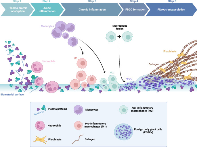
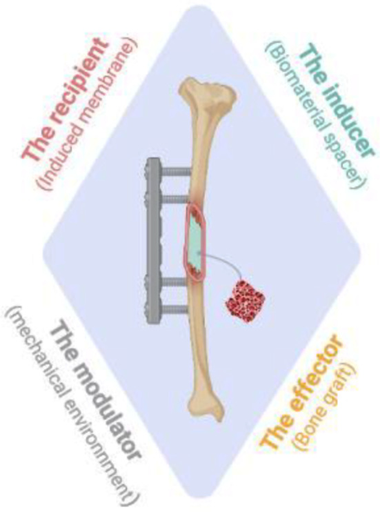
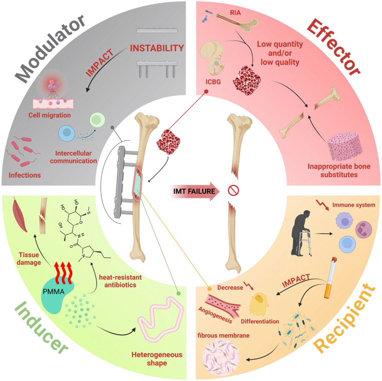
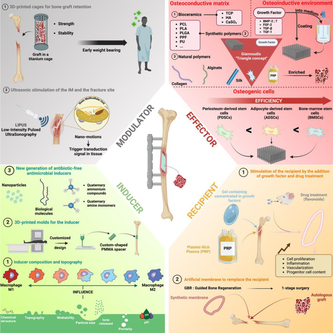
The reconstruction of large bone defects remains challenging for orthopedic surgeons. Autologous bone grafts (ABGs) are the gold standard treatment for limited size defects, but larger bone defects (> 5 cm) require the use of more sophisticated techniques, such as the Masquelet technique. Over the last three decades, the Masquelet or induced membrane technique (IMT) has become increasingly popular as it does not require high-precision microsurgery skills and the time taken to achieve bone consolidation is independent of the length of the defect. IMT is a two-stage procedure. In the first stage, a polymethylmethacrylate (PMMA) cement spacer is implanted into the bone lesion and a physiological immune reaction initiates the formation of a fibrotic induced membrane (IM) with both angiogenic and osteogenic properties. The second stage, performed several weeks later, involves removal of the spacer followed by the implantation of a standard ABG in the preserved IM cavity for subsequent bone repair. In this extensive review, we explain how the success of this surgical procedure can be attributed to the synergy of four key components: the inducer (the PMMA cement), the recipient (the IM), the effector (the bone graft) and the modulator (the mechanical environment). Conversely, we then explain how each key component can contribute to the failure of such treatment. Finally, we discuss existing or emerging innovative and biotechnology-oriented strategies for optimizing surgical outcome with respect to the four components of IMT described above.

Keywords: Bone repair; Foreign body reaction; Induced membrane; Masquelet; Orthopedic surgery.


Conflict of interest statement

Declarations. Competing interests: The authors declare no competing interests.

Figures

Fig. 1
Fig. 1
The foreign-body reaction underlying induced-membrane formation. After PMMA spacer implantation, plasma protein adsorption occurs rapidly at the cement surface (step 1), triggering neutrophil mobilization, which initiates an acute inflammatory response (step 2). An infiltration of lymphocytes and monocytes then occurs, followed by monocyte differentiation into macrophages over a period of several days, corresponding to the chronic inflammation response (step 3). In the next step (step 4), macrophages are activated and fused into foreign body giant cells (FBGC). The formation of these cells is a physiological attempt to eliminate the PMMA cement through phagocytosis. Finally (step 5), fibroblasts are recruited and activated to produce a collagen-based extracellular matrix (ECM) involved in the formation of a fibrous capsule to isolate the PMMA cement from the patient’s tissues. Created with BioRender.com
Fig. 2
Fig. 2
Representative organization of a human induced membrane stained with Masson-Goldner trichrome dye. The black asterisk outside the section indicates the location of the PMMA spacer responsible for membrane induction. Three different layers are identified in the induced membrane (indicated by yellow letters). a: is the “inner” layer closest to the spacer consisting principally of monocyte-derived cells. b: is the “intermediate” layer containing small blood vessels, fibroblast-like cells and collagen fibers orientated in parallel to the PMMA spacer. c: is the “external” layer characterized by unorganized collagen fibers, a combination of myofibroblasts, fibroblast-like cells and large blood vessels. The scale bar represents 50 μm
Fig. 3
Fig. 3
The concept underlying the induced-membrane technique. The success of the procedure depends on the interaction between an inducer (the spacer), a recipient (the IM) and an effector (the graft). The mechanical environment of the fracture acts as a modulator of these components. Created with BioRender.com
Fig. 4
Fig. 4
Overview of the principal factors associated with a poor outcome of IMT. According to the induced-membrane concept illustrated in Fig. 3, various factors may lead to failure of the induced-membrane technique (IMT). Failures related to the “inducer” component generally result from several drawbacks of the PMMA spacer itself, such as the heat generated by PMMA cement polymerization. The “recipient” component may be adversely affected by an unhealthy patient lifestyle, such as smoking, or old age. For the “effector” component, a small amount of autologous bone graft material or the poor quality of the graft may decrease bone repair efficiency. The use of an inappropriate bone substitute may also have a similar effect. Finally, the instability of the osteosynthesis system (bone fixators) provides an example of the contribution of the “modulator” component to IMT failure. Created with BioRender.com
Fig. 5
Fig. 5
Overview of promising strategies for improving the therapeutic outcome of the induced-membrane technique. This figure summarizes the most promising medical and technological strategies for increasing the bone-repair efficiency of the induced-membrane technique taking into account the inducer, recipient, effector and modulator aspects of the Fig. 3 IMT concept. Created with BioRender.com

Similar articles

Cited by

References

    1. Greenwald AS, et al. Bone-graft substitutes: facts, fictions, and applications. J Bone Joint Surg Am 83-A Suppl 2 Pt. 2001;2:98–103. - PubMed
    1. Cuthbert RJ, et al. Induced periosteum a complex cellular scaffold for the treatment of large bone defects. Bone. 2013;57:484–92. - PubMed
    1. Klopfleisch R, Jung F. The pathology of the foreign body reaction against biomaterials. J Biomed Mater Res A. 2017;105:927–40. - PubMed
    1. Sheikh Z, Brooks PJ, Barzilay O, Fine N, Glogauer M, Macrophages. Foreign body giant cells and their response to implantable biomaterials. Mater (Basel). 2015;8:5671–701. - PMC - PubMed
    1. Trindade R, Albrektsson T, Tengvall P, Wennerberg A. Foreign body reaction to biomaterials: on mechanisms for buildup and breakdown of osseointegration. Clin Implant Dent Relat Res. 2016;18:192–203. - PubMed

MeSH terms

LinkOut - more resources