Spatial Heterogeneity of PD-L1 Expression as a Biomarker for Third-Generation EGFR-TKI Response in Advanced EGFR-Mutant NSCLC
- PMID: 40102299
- PMCID: PMC12127114
- DOI: 10.1111/cas.70060
Spatial Heterogeneity of PD-L1 Expression as a Biomarker for Third-Generation EGFR-TKI Response in Advanced EGFR-Mutant NSCLC
Abstract
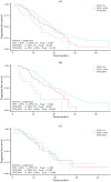
The association between the spatial heterogeneity of programmed cell death ligand 1 (PD-L1) expression and the efficacy of third-generation epidermal growth factor receptor tyrosine kinase inhibitors (EGFR-TKIs) in EGFR-mutant non-small cell lung cancer (NSCLC) remains elusive. This retrospective study analyzed data from 4171 NSCLC patients with EGFR-sensitive mutations treated at Shanghai Chest Hospital from August 2019 to September 2023. Among them, 182 patients receiving third-generation EGFR-TKIs monotherapy as a first-line treatment were enrolled. Patients were categorized by biopsy sites into primary lung lesions (n = 112) and metastatic lymph nodes (n = 70). PD-L1 expression was stratified based on tumor cell proportion score (TPS): < 1%, 1%-49%, and ≥ 50%. The median progression-free survival (PFS) for the entire cohort was 18.33 months. In the PD-L1 TPS group, PFS was 18.87 months for TPS < 1%, 17.6 months for TPS 1%-49%, and 13.6 months for TPS ≥ 50%, with significant differences across groups (p = 0.026). Moreover, multivariate analysis identified smoking history [HR = 1.653, 95% CI (1.132-2.414), p = 0.009] and TPS ≥ 50% [HR = 2.069, 95% CI (1.183-3.618), p = 0.011] as independent risk factors. In primary lesions, the median PFS was 21.93 months for TPS < 1%, 18.57 months for TPS 1%-49%, and 10.17 months for TPS ≥ 50%, with significant differences (p < 0.001). However, PD-L1 expression in metastatic lymph nodes was not associated with PFS (p = 0.973). In advanced EGFR-mutant NSCLC, high PD-L1 expression may suggest reduced efficacy of third-generation EGFR-TKIs. The spatial heterogeneity of PD-L1 expression could influence its predictive accuracy for third-generation EGFR-TKI efficacy.
Keywords: EGFR; NSCLC; PD‐L1; heterogeneity; third‐generation EGFR‐TKIs.
© 2025 The Author(s). Cancer Science published by John Wiley & Sons Australia, Ltd on behalf of Japanese Cancer Association.
Conflict of interest statement
The authors declare no conflicts of interest.
Figures


Similar articles
-
Heterogeneity in PD-L1 expression between primary and metastatic lymph nodes: a predictor of EGFR-TKI therapy response in non-small cell lung cancer.Respir Res. 2024 Jun 5;25(1):233. doi: 10.1186/s12931-024-02858-3. Respir Res. 2024. PMID: 38840238 Free PMC article.
-
[A Real-world Study on the Expression Characteristics of PD-L1 in Patients with Advanced EGFR Positive NSCLC and Its Relationship with the Therapeutic Efficacy of EGFR-TKIs].Zhongguo Fei Ai Za Zhi. 2023 Mar 20;26(3):217-227. doi: 10.3779/j.issn.1009-3419.2023.101.09. Zhongguo Fei Ai Za Zhi. 2023. PMID: 37035884 Free PMC article. Chinese.
-
Strong PD-L1 affect clinical outcomes in advanced NSCLC treated with third-generation EGFR-TKIs.Future Oncol. 2024;20(32):2481-2490. doi: 10.1080/14796694.2024.2385290. Epub 2024 Aug 19. Future Oncol. 2024. PMID: 39155845 Free PMC article.
-
Epidermal Growth Factor Receptor (EGFR) Pathway, Yes-Associated Protein (YAP) and the Regulation of Programmed Death-Ligand 1 (PD-L1) in Non-Small Cell Lung Cancer (NSCLC).Int J Mol Sci. 2019 Aug 5;20(15):3821. doi: 10.3390/ijms20153821. Int J Mol Sci. 2019. PMID: 31387256 Free PMC article. Review.
-
PD-L1 expression and EGFR status in advanced non-small-cell lung cancer patients receiving PD-1/PD-L1 inhibitors: a meta-analysis.Future Oncol. 2019 May;15(14):1667-1678. doi: 10.2217/fon-2018-0639. Epub 2019 May 1. Future Oncol. 2019. PMID: 31041879
References
MeSH terms
Substances
Grants and funding
LinkOut - more resources
Full Text Sources
Medical
Research Materials
Miscellaneous

