. 2025 Mar 18;16(1):184.
doi: 10.1038/s41419-025-07516-0.

Monounsaturated fatty acids promote cancer radioresistance by inhibiting ferroptosis through ACSL3

Affiliations

Monounsaturated fatty acids promote cancer radioresistance by inhibiting ferroptosis through ACSL3

Yulin Cao et al. Cell Death Dis. .

Erratum in

Abstract

Radioresistance is a major challenge in tumor radiotherapy and involves in a mixture of cellular events, including ferroptosis, a new type of programmed cell death characterized by the excess accumulation of iron-dependent lipid peroxides. In the present study, we observed that surviving cancer tissues and cells after radiotherapy had significantly greater glutathione to oxidized glutathione (GSH/GSSG) ratios and lower lipid reactive oxygen species (ROS) and malondialdehyde (MDA) levels than nonirradiated tumors and cells. Untargeted lipidomic analyses revealed that oleic acid (OA) and palmitoleic acid (POA) were the most significantly upregulated unsaturated fatty acids in irradiated surviving cancer cells compared with those in control cancer cells irradiated with IR. Both OA and POA could protect cancer cells from the killing effects of the ferroptosis inducer erastin and RSL3, and OA had a stronger protective effect than POA, resulting in lower lipid ROS production than POA. Mechanistically, OA protected cells from ferroptosis caused by the accumulation of polyunsaturated fatty acid-containing phospholipids in an ACSL3-dependent manner. A mouse model demonstrated that ACSL3 knockdown combined with imidazole ketone erastin synergistically enhanced antitumor effects in radiation-resistant tumors in vivo. Our study reveals previously undiscovered associations between radiation and fatty acid metabolism and ferroptosis, providing a novel treatment strategy for overcoming cancer radioresistance.


Conflict of interest statement

Competing interests: The authors declare no competing interests. Ethics: The present study was approved by the Clinical Research Ethics Committees of the participating institutions. Human primary rectal cancer tissues from patients receiving neoadjuvant RT (before and after RT) were collected from Affiliated Hospital of Jiangnan University with informed consent. The study was approved by the Clinical Research Ethics Committees of Affiliated Hospital of Jiangnan University [LS2021104]. All animal experiments were performed in accordance with the relevant institutional and national guidelines and the regulations of Jiangnan University Medical Experimental Animal Care Commission (JN. No20230228c0480515).

Figures
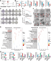
Fig. 1
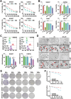
Fig. 1. Decreased ferroptosis contributes to radioresistance in radioresistant cancer cells.
A Treatment schedules for the CMT93 (n = 3) and A549 (n = 3) subcutaneous xenograft models, which were exposed to 8 Gy or 12 Gy of X-rays, respectively. B Quantitative analyses of lipid peroxidation and GSH/GSSG levels in nonirradiated control tumors and radioresistant tissues (RR) after RT (n = 3). C, D Representative images of clonogenic survival assays of CMT93 or CMT93-RR cells subjected to 0 to 28 Gy of X-ray, SW837 or SW837-RR cells subjected to 0 to 22 Gy of X-ray. The dose survival curves were plotted using GraphPad Prism 9.0 software. E Heatmap of differentially expressed proteins between radioresistant and control cells involved in ferroptosis. F TEM images of cell morphology after IR treatment (12 Gy for SW837, 22 Gy for CMT93) for 24 h; red arrows indicate mitochondria. G Quantitative analyses of lipid peroxidation and GSH/GSSG levels in reirradiated radioresistant cancer cells and control cells. (n = 3) *p < 0.05, **p < 0.01, ***p < 0.001, ****p < 0.0001.
Fig. 2
Fig. 2. IR treatment alters the FA metabolism profiles of radioresistant cancer cells.
HDMB (A) and KEGG (B) analyses show the changes in fatty acid metabolism in nonirradiated cancer cells, irradiated cancer cells, and their corresponding radioresistant cells. C Flow chart showing the results of the intersection of significantly changed unsaturated FAs in nonirradiated control cells, cancer cells treated with IR, SW837-RR and CMT93-RR cells.
Fig. 3
Fig. 3. OA protects radioresistant cells from ferroptosis.
A The cell cytotoxicity of different concentrations (0-100 nM) of OA and POA to SW837 and CMT93 cells at 12 h, 24 h, and 36 h (n = 3). B CCK-8 assays were performed to determine the effects of erastin (1 μM), OA (20 nM), and POA (20 nM) on the viability of SW837, CMT93, A549, and H1299 cells (n = 3). C Quantitative analysis of lipid peroxidation levels and the GSH/GSSG ratio in SW837, CMT93, A549, and H1299 cells treated with 1 μM erastin, 20 nM OA, or 20 nM POA (n = 3). D Cell morphology was observed via TEM after cells were treated with or without OA (20 nM) for 24 h, and then treated with IR (12 Gy for SW837, 22 Gy for CMT93) for 24 h; red arrows, mitochondria. E Effect of OA pretreatment on colony formation. The cells were plated in 6-well plates for 12 h and then irradiated with 0 to 28 Gy of X-ray radiation using a linear accelerator. The cells were grown at 37 °C for 14 days, after which the number of colonies containing 50 or more cells was counted. Each experiment was performed at least three times. F Dose survival curves of CMT93 or CMT93-RR cells subjected to 0–28 Gy of X-ray irradiation, and SW837 or SW837-RR cells subjected to 0–22 Gy of X-ray irradiation. *p < 0.05, **p < 0.01, ***p < 0.001, ****p < 0.0001.
Fig. 4
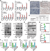
Fig. 4. Ferroptosis resistance in radioresistant cancer cells depends on ACSL3.
A The mRNA expression of ferroptosis-related genes in radioresistant cancer cells (SW837-RR, CMT93-RR, A549-RR, H1299-RR) and their parental cells (SW837, CMT93, A549, H1299). β-actin was used as an internal control (n = 3). B The protein levels of ACSL3, GPX4, Nrf2, SLC7A11, and TfR1 in radioresistant cancer cells were measured by Western blotting. IHC staining (C) and the scores (D) of ACSL3 protein expression in paired nonirradiated and irradiated tumor tissue samples from rectal cancer patients (n = 24). The upper scale bar represents 200 μm. The lower scale bar represents 100 μm. E The knockout efficiency of ACSL3 in SW837-RR and CMT93-RR cells was determined by western blotting. F CCK-8 assays were performed to determine the effects of 1 μM erastin on the viability of ACSL3-KO SW837-RR and CMT93-RR cells (n = 3). G Lipid peroxidation levels in ACSL3 KO radioresistant cancer cells. These cells were treated with DMSO (control) or 1 μM erastin (n = 3). H Quantitative analyses of GSH/GSSG ratio in ACSL3 KO radioresistant cancer cells. These cells were treated with DMSO (control) or 1 μM erastin (n = 3). *p < 0.05, **p < 0.01, ***p < 0.001.
Fig. 5
Fig. 5. OA protects radioresistant cells from ferroptosis depending on ACSL3.
A Viability of sgACSL3-1, sgACSL3-2 SW837-RR and CMT93-RR cells treated with or without 1 μM erastin or 20 nM OA exposure. Error bars are the means ± SD (n = 3). B Clonogenic survival assays were performed to evaluate the colony formation ability of ACSL3-KO or control cancer cells after treatment with 1 μM erastin or 20 nM OA. Student’s t-tests were used. Quantitative analyses of lipid peroxidation levels (C) and the GSH/GSSG ratio (D) in sgACSL3-1, sgACSL3-2 SW837-RR and CMT93-RR cells treated with or without 1 μM erastin or 20 nM OA. Lipid levels determined by mass spectrometry in SW837, SW837-RR, and SW837-RR-ACSL3 KO cells (E) or CMT93, CMT93-RR, and CMT93-RR-ACSL3 KO cells (F). PE phosphatidylethanolamine, PC phosphatidylcholine, PS phosphatidylserine. *p < 0.05, **p < 0.01, ***p < 0.001, ****p < 0.0001.
Fig. 6
Fig. 6. Lipid droplet (LD) formation is not required for OA to inhibit ferroptosis.
A Oil red O staining of ACSL3 KO SW837-RR and CMT93-RR cells with or without 20 nM OA exposure. Scale bars, 50 μm. B The overexpression efficiency of ACSL3 in SW837 and CMT93 cells was determined by western blotting. C Images of Oil Red O stained ACSL3-OE SW837 and CMT93 cells exposed to DGATis (a combination of T863 (20 μM) and PF-06424439 (10 μM)). D CCK-8 assays were performed to determine the effects of 20 nM OA and DGATis (a combination of T863 (20 μM) and PF-06424439 (10 μM)) on the viability of ACSL3-OE SW837 and CMT93 cells. Quantitative analyses of lipid peroxidation levels (E) and the GSH/GSSG ratio (F) in OE ACSL3 SW837 and CMT93 cells treated with or without OA (20 nM) and DGATis. *p < 0.05, **p < 0.01, ***p < 0.001, ****p < 0.0001.
Fig. 7
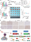
Fig. 7. shACSL3-AAV combined with IKE significantly represses radioresistance in vivo.
A CCK-8 assays were performed to determine the effects of 50 µM IKE and shACSL3-AAV (450,000 genome copies/cell) on the viability of CMT93-RR cells. B The treatment schedule for the CMT93-RR syngeneic mouse model. shACSL3-AAV was administered intratumorally at a dose of 1.8 × 1011 viral particles per animal. Injections were performed once every two days for a total of 6 times. Mice were treated with PBS, 30 mg/kg IKE, or shACSL3-AAV (1.8 × 1011 viral particles per animal) as a single agent or in combination. C Representative images of tumors. Tumor volume (D), tumor weights (E), and body weights of the mice (F). (n = 5) G The expression Ki67 and 4-HNE in the different treatment groups was measured by IHC. H Schematic illustration demonstrating that OA regulates ferroptosis and radioresistance in an ACSL3-dependent manner. *p < 0.05, **p < 0.01, ***p < 0.001, ****p < 0.0001.

References

    1. Ye LF, Chaudhary KR, Zandkarimi F, Harken AD, Kinslow CJ, Upadhyayula PS, et al. Radiation-induced lipid peroxidation triggers ferroptosis and synergizes with ferroptosis inducers. ACS Chem Biol. 2020;15:469–84. - PMC - PubMed
    1. Yuan Z, Liu T, Huo X, Wang H, Wang J, Xue L. Glutamine transporter SLC1A5 regulates ionizing radiation-derived oxidative damage and ferroptosis. Oxid Med Cell Longev. 2022;2022:3403009. - PMC - PubMed
    1. Chen X, Kang R, Kroemer G, Tang D. Broadening horizons: the role of ferroptosis in cancer. Nat Rev Clin Oncol. 2021;18:280–96. - PubMed
    1. Gebicki JM, Du J, Collins J, Tweeddale H. Peroxidation of proteins and lipids in suspensions of liposomes, in blood serum, and in mouse myeloma cells. Acta Biochim Pol. 2000;47:901–11. - PubMed
    1. Spitz DR, Azzam EI, Li JJ, Gius D. Metabolic oxidation/reduction reactions and cellular responses to ionizing radiation: a unifying concept in stress response biology. Cancer Metastasis Rev. 2004;23:311–22. - PubMed

MeSH terms