. 2025 Mar 18;16(1):2676.
doi: 10.1038/s41467-025-57980-7.

Global characterization of mouse testis O-glycoproteome landscape during spermatogenesis

Affiliations

Global characterization of mouse testis O-glycoproteome landscape during spermatogenesis

Qiannan Liu et al. Nat Commun. .

Abstract

Protein O-glycosylation plays critical roles in sperm formation and maturation. However, detailed knowledge on the mechanisms involved is limited due to lacking characterization of O-glycoproteome of testicular germ cells. Here, we performed a systematic analysis of site-specific O-glycosylation in mouse testis, and established an O-glycoproteome map with 349 O-glycoproteins and 799 unambiguous O-glycosites. Moreover, we comprehensively investigated the distribution properties of O-glycosylation in testis and identified a region near the N-terminal of peptidase S1 domain that is susceptible to O-glycosylation. Interestingly, we found dynamic changes with an increase Tn and a decrease T structure from early to mature developmental stages. Notably, the importance of O-glycosylation was supported by its effects on the stability, cleavage, and interaction of acrosomal proteins. Collectively, these data illustrate the global properties of O-glycosylation in testis, providing insights and resources for future functional studies targeting O-glycosylation dysregulation in male infertility.


Conflict of interest statement

Competing interests: The authors declare no competing interests.

Figures
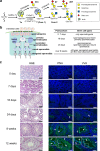
Fig. 1
Fig. 1. Cellular localization of O-GalNAc glycans in mouse testes at different ages.
a Illustration of the major O-GalNAc glycans. Biosynthesis and extension of Tn and T structures were shown. b Schematic diagram of different stages of spermatogenesis. The spermatogonia perform a mitotic division to form primary spermatocytes. The latter undergo two meiotic divisions to form round spermatids, which subsequently undergo a structural metamorphosis to become elongated spermatids. The composition of germ cell types in seminiferous tubules of mouse testes from 0 day after birth until sexual maturity was summarized according to literature. c H&E (Hematoxylin and Eosin) staining (left) and lectin staining (middle and right) of seminiferous tubules of mouse testes at different times after birth. The signal of PNA (peanut agglutinin) recognizing T antigen (Galβ1,3GalNAc-α1-Ser/Thr) was strongly stained in round and elongated spermatids, especially in round spermatids. The signal of VVA (vicia villosa agglutinin) recognizing Tn antigen (GalNAc-α1-Ser/Thr) was strongly stained in round and elongated spermatids, especially in elongated ones. White hollow and solid arrows indicate round and elongated spermatids, respectively. The images shown are representative of five mice/group. Scale bars represent 50 μm. STn sialylated Tn antigen, ST sialylated T antigen, dST di-sialylated T antigen.
Fig. 2
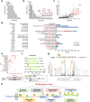
Fig. 2. In-depth characterization of the O-glycoproteome in testes of 24-day-old and 12-week-old mice.
a Schematic workflow for analyzing the testis O-glycoproteome. Proteins from testes of 24-day-old and 12-week-old mice were extracted, digested by trypsin, and subsequently de-sialylated using neuraminidase. The O-glycopeptides with Tn and T structures were enriched using VVA and PNA lectins respectively, and then identified by LC-MS/MS using higher-energy dissociation product ions-triggered electron-transfer/higher-energy collision dissociation (HCD-pd-EThcD) strategy. Four biological replicates were used for each group. Illustrations used elements from Servier Medical Art (http://smart.servier.com/) under a Creative Common Attribution 3.0 Generic License (https://creativecommons.org/licenses/by/3.0/). b The numbers of O-glycoproteins, unique O-glycopeptides, unambiguous O-glycosites, and glycopeptide-spectrum matches (GPSM) identified in each group. c Venn diagram showing the distribution of identified O-glycoproteins, O-glycopeptides, and O-glycosites enriched from testes at different developmental stages using different lectins. d, e Overlap of the testis O-glycoproteins identified in this study with those from the previously reported mouse O-glycoproteins atlas (d), as well as with the reported O-glycoproteins of the other 10 mouse organs published by Yang et al.. (e, f) Overlap of human counterparts of the 337 mouse O-glycoproteins with the published human O-glycoproteins which were mainly identified by chemical methods, LWAC or EXoO strategy. VVA vicia villosa agglutinin, PNA peanut agglutinin, Tn GalNAc-α1-Ser/Thr, T Galβ1,3GalNAc-α1-Ser/Thr, GPSM glycopeptide-spectrum matches, LWAC lectin weak affinity chromatography, EXoO site-specific extraction of O-linked glycopeptides.
Fig. 3
Fig. 3. Density mapping and distribution preference of O-glycosites in mouse testes.
a, b The number of unambiguous O-glycosites per peptide (a) and per protein (b). Glycoproteins involved in spermatogenesis and fertilization were labeled red. c The number of unique O-glycopeptides and GPSM per protein. The glycoproteins with a high number of glycopeptides (≥ 5) and high abundance of GPSM (> 100) were labeled in red. d Twenty glycopeptides with dense O-glycosites (≥ 5) were listed, and the tissue specificity of the corresponding proteins was shown with reference to the UniProt database. The histogram shows the number of GPSM of these glycopeptides identified in each group. e The protein domains containing O-glycosites located at the outer or inner edges (≥  2 sites) were shown. The domain edge is defined as 20 amino acids located inside (inner edge, positive) or outside (outer edge, negative) the N-/C-terminal of the domain. f A region near the N-terminal of domain peptidase S1 domain was found to be susceptible to O-glycosylation. The glycosites identified in testes of 24-day-old and 12-week-old mice are indicated by yellow and orange boxes, respectively. g An illustrative annotated MS2 spectrum of the O-glycopeptide from PRSS43 modified with T structure [Hex(1)HexNAc(1)] was shown. h Summary of distribution percentage of 799 O-glycosites identified in mouse testes. Protein annotations were referred against the UniProt database. GPSM glycopeptide-spectrum matches, VVA vicia villosa agglutinin, PNA peanut agglutinin. Source data are provided as a Source Data file.
Fig. 4
Fig. 4. Quantitative analysis of O-glycopeptides identified in mouse testes at different stages of spermatogenesis.
a Unsupervised hierarchical clustering analysis of O-glycopeptides enriched with VVA or PNA lectin from 24-day-old and 12- week-old testes. The label-free quantitative data of O-glycopeptides were obtained using pGlycoQuant. b The distribution of glycan compositions on unambiguous O-glycosites identified in 24-day-old and 12-week-old testes. The number and percentage of GPSM corresponding to H1N1 and N1 glycans were shown. c, d Volcano plots of individual O-glycopeptide abundance fold changes (log2 scale) and corresponding p-values (-log10 scale) enriched by VVA (c) or PNA (d). The p-value was calculated by two-tailed Student’s t-test. Up-regulated and down-regulated glycopeptides ( with > 2-fold changes and p < 0.05) were highlighted in red and blue, respectively, and the top 10 glycopeptides in each group having the lowest p-values were labeled. e Gene Ontology (GO) biological process terms enriched in up-regulated and down-regulated glycoproteins in testes of 12-week-old mice. Significance was calculated by one-tailed Fisher’s Exact Test (Benjamini-Hochberg FDR-adjusted p < 0.05). The top 10 up-regulated (red) and down-regulated (blue) terms with the lowest p-values were shown. f, g Differential glycoproteins involving in fertilization were depicted. The unambiguous O-glycosites (f) and the number of GPSM (g) of the 13 glycoproteins with altered abundance between testes of 24-day-old and 12-week-old mice were shown. VVA vicia villosa agglutinin, PNA peanut agglutinin, H Hexose, N N-acetylhexosamine, GPSM glycopeptide-spectrum matches. Source data are provided as a Source Data file.
Fig. 5
Fig. 5. O-glycosylation patterns of round and elongated spermatids from 24-day and 12-week testes.
ad Schematic representation of the flow cytometry gating strategy used to sort haploid spermatids from 24-day-old (a) and 12-week-old testes (c). Cell debris and doublets were first excluded. Subsequently, Hoechst-stained cells were visualized on a Hoechst-blue/Hoechst-red contour plot, and haploid cells with 1C DNA content were gated. The round and elongated spermatids were further distinguished using two imaging parameters (eccentricity and short axis moment). The sorted round spermatids from 24-day testes (b), as well as round and elongated spermatids from 12-week testes (d), were visualized by BD CellViewTM imaging. eg VVA blot (e), PNAblot (f), and silver staining (g) of sorted round and elongated spermatids proteins from 24-day-old (n = 20) and 12-week-old (n = 10) mice. The number of mice used in each batch  was shown. h, i Molecular weight (Mw) distributions of up-regulated glycoproteins in the VVA-enriched subset (h) and down-regulated glycoproteins in the PNA-enriched subset (i) in 12-week-old testicular tissues identified by mass spectrometry (MS). The Mw value shown in (h, i) represents the theoretical molecular weight of the protein plus the molecular weight of the O-glycans identified in this study. VVA vicia villosa agglutinin, PNA peanut agglutinin. Source data are provided as a Source Data file.
Fig. 6
Fig. 6. O-glycosylation affects interaction of pro-ACR and ACRBP.
a Protein schematic representation illustrating the location of O-glycosites on pro-ACR and ACRBP. The schematic positions and sequences of six synthetic peptides of pro-ACR and ACRBP were shown. b Sankey diagrams showing site-specific O-glycan changes in the testis from 24 days to 12 weeks. The number of glycopeptide-spectrum matches corresponding to each glycan composition was reflected in the thickness of the flow line and the labeled number on the rectangle. c, d Representative MS2 spectra showing the O-glycosites and O-glycan structures in glycopeptides of pro-ACR (c) and ACRBP (d). e Flag-tagged pro-ACR or His-tagged ACRBP was transfected into CHO-K1 or CHO-ldlD cells. The VVA signal showing decreased levels of O-GalNAc glycan on proteins expressed in CHO-ldlD cells. f Co-Immunoprecipitation analysis of pro-ACR and ACRBP. Flag-tagged pro-ACR and His-tagged ACRBP were co-transfected into CHO-K1 or CHO-ldlD cells. Anti-Flag or anti-His antibody was used to detect the expression of pro-ACR or ACRBP. GAPDH was used as a loading control. g Chromatograms of glycosylated products of pro-ACR and ACRBP peptides catalyzed by O-glycosyltransferases GALNT3, GALNT1, and GALNTL5, respectively. The peptides localized in the region where pro-ACR interacts with ACRBP were labeled red. Data in (e, f) are representative of two or three independent experiments. H Hexose, N N-acetylhexosamine, F Fucose, VVA vicia villosa agglutinin. Source data are provided as a Source Data file.
Fig. 7
Fig. 7. O-glycosylation functions on SPACA7, INSL6, EQTN, and SPESP1.
The Flag-EGFP-tagged SPACA7 (a), INSL6 (c), EQTN (e), and SPESP1 (g) were transfected into CHO-K1 or CHO-ldlD cells. The VVA signals showing decreased levels of O-GalNAc glycan on proteins expressed in CHO-ldlD cells. (b) The role of O-glycosylation in protein stability was assessed. CHO cells were treated with 100 μg/mL CHX, and cell lysates were collected at various time points. The intensity of SPACA7 was normalized to that of GAPDH, and the percentage of remaining SPACA7 at different time points was calculated. d The role of O-glycosylation in protein maturation and cleavage was evaluated. The cell lysate and culture supernatant from the same batch of CHO cells were collected. The expression of pro-INSL6 and cleavage peptides were detected using anti-Flag antibody. f The role of O-glycosylation in protein-protein interaction was investigated. Co-Immunoprecipitation analysis was performed for EQTN and SNAP25. Flag-EGFP-tagged EQTN and His-EGFP-tagged SNAP25 were co-transfected into CHO-K1 or CHO-ldlD cells. Anti-Flag or anti-His antibody was used to detect the expression of EQTN or SNAP25. h The role of O-glycosylation in protein dimer formation was examined. Western blot analysis of cell lysates from the CHO cells transfected with SPESP1 under non-denatured or denatured conditions was detected by anti-Flag antibody. Data in (ah) are representative of two or three independent experiments. VVA vicia villosa agglutinin, CHX cycloheximide, M marker, CBB Coomassie Brilliant Blue stain. Source data are provided as a Source Data file.

Similar articles

References

    1. Eisenberg, M. L. et al. Male infertility. Nat. Rev. Dis. Prim.9, 49 (2023). - PubMed
    1. Tatum, M. China’s fertility treatment boom. Lancet396, 1622–1623 (2020). - PubMed
    1. Agarwal, A. et al. Male infertility. Lancet397, 319–333 (2021). - PubMed
    1. Auger, J. Spermatogenic Cells—Structure. Encycl. Reprod.1, 53–60 (2018).
    1. Tecle, E. & Gagneux, P. Sugar-Coated Sperm: Unraveling the Functions of the Mammalian Sperm Glycocalyx. Mol. Reprod. Dev.82, 635–650 (2015). - PMC - PubMed

LinkOut - more resources