. 2025 Mar 18;16(1):2682.
doi: 10.1038/s41467-025-57944-x.

The primate-specific Nedd4-1(NE) localizes to late endosomes in response to amino acids to suppress autophagy

Affiliations

The primate-specific Nedd4-1(NE) localizes to late endosomes in response to amino acids to suppress autophagy

G Kefalas et al. Nat Commun. .

Abstract

The ubiquitin ligase Nedd4 (Nedd4-1), comprised of C2-WW(n)-HECT domains, regulates protein trafficking. We recently described a primate-specific Nedd4-1 splice isoform with an extended N-terminus replacing the C2 domain, called Nedd4-1(NE). Here, we show that while canonical Nedd4-1 is primarily localized to the cytosol, Nedd4-1(NE) localizes to late endosomes. This localization is mediated by the NE region, is dependent on amino acid availability, is independent of mTORC1, and is inhibited by the autophagy inducer IKKβ. We further demonstrate that VPS16B, which regulates late endosome to lysosome maturation, is a unique Nedd4-1(NE) substrate that co-localizes with Nedd4-1(NE) in the presence of nutrients. Importantly, a potentially pathogenic homozygous variant identified in the NE region (E70Q) of a patient with lymphangiectasia and protein-losing enteropathy leads to reduced VPS16B ubiquitination by Nedd4-1(NE). Finally, we report that Nedd4-1(NE) inhibits autophagy, likely by disrupting late endosome to autophagosome maturation. This work identified an mTORC1-independent, IKK-driven mechanism to regulate Nedd4-1(NE) localization to late endosomes in primates in response to nutrient availability, and uncovered suppression of autophagy by this ubiquitin ligase.


Conflict of interest statement

Competing interests: The authors declare no competing interests.

Figures

Fig. 1
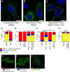
Fig. 1. Nedd4-1(NE) is localized to late endosomes.
A Schematic representation of the canonical Nedd4-1 and the Nedd4-1(NE) isoform. Due to alternative splicing, Nedd4-1(NE) contains a 516-amino acid NE region and only a small part of the C2 domain. Images are drawn to scale. BE HeLa cells were transfected with Flag-tagged constructs, as indicated. Cells were fixed, permeabilized, and immunostained with anti-Flag antibodies (green). Nuclei were stained with DAPI (blue). F HeLa cells were treated as in (B), and fixed samples were additionally immunostained using antibodies against endogenous Rab7 (red). G Quantification of the percentage of overlap of Nedd4-1(NE) with various endogenous protein markers. Data are means ± SD (N = 3 independent experiments, n = 27-36 cells/experiment; precise values are indicated in the Source Data file). All scale bars are 10 μm.
Fig. 2
Fig. 2. Nedd4-1(NE) localization is regulated by essential amino acids.
HeLa cells were transfected with 3xFlag-Nedd4-1(NE) and incubated for 1 hr in (A) DMEM (supplemented with 10% serum), (B) HBSS, or (C) HBSS supplemented with an essential amino acid (EAA) cocktail plus glutamine (Gln; 4 mM). Cells were fixed, permeabilized, and immunostained with anti-Flag antibodies (green). Nuclei were stained with DAPI (blue). DJ Nedd4-1(NE) localization was scored blindly as either “diffuse - cytoplasm/membrane”, “punctate”, or “both”, as indicated by the examples in (HJ). Scale bars are (AC) 10 μm, or (HJ) 15 μm. Data are means ± SEM of 3 independent experiments. Statistical significance was determined using a chi-squared test (N.S.: not significant; **: p < 0.01; ****: p < 0.0001). In (E), statistical tests are comparing each condition to HBSS. Cell counts for each condition are indicated in Table S2.
Fig. 3
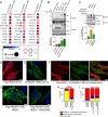
Fig. 3. IKKβ inhibits Nedd4-1(NE) localization to late endosomes.
A 293 T-REx cells expressing 3xFlag-miniTurbo (FmT)-tagged constructs of Nedd4-1 or Nedd4-1(NE) were treated with biotin for 30 min and then collected. Biotinylated proteins in the lysates were identified by mass spectrometry. All raw mass spectrometry data files have been deposited at the MassIVE archive (massive.ucsd.edu), accession ID MSV000093801. Proteins involved in autophagy and protein trafficking were identified using GO-TermFinder and dot plots were generated using ProHits-viz web tool. Prey abundance across baits and prey confidence are displayed as indicated in the legend (below). B HEK293T cells were transfected with the indicated constructs. Cell lysates were subjected to Flag immunoprecipitation (IP), and the indicated proteins were detected by immunoblotting. Constructs labeled “CS” correspond to full-length, catalytically inactive (Cys → Ser) mutants. C HEK293T cells were transfected with Flag-NE and incubated in the absence or presence of BI605906 (10 μM, 2 hrs) and Calyculin A (50 nM, 10 min). Cell lysates were incubated in 1% SDS and boiled to dissociate non-covalent protein complexes prior to Flag IP. NE phosphorylation was detected by immunoblotting. The markers beside each blot indicate molecular weight (measured in kDa). DG HeLa cells were transfected with (D) Flag-IKKβ alone, (E), V5-Nedd4-1(NE) alone, (F) both Flag-IKKβ and V5-Nedd4-1(NE), or (G) 3xFlag-Nedd4-1(NE) alone. Cells were incubated in (DF) DMEM, or (G) HBSS, in the absence or presence of 10 μM BI605906, for 2 hrs. Cells were fixed, permeabilized, and immunostained with (DF) anti-Flag (IKKβ, red) and/or anti-V5 (Nedd4-1(NE), green) antibodies, or (G) anti-Flag (Nedd4-1(NE), green). Nuclei were stained with DAPI (blue). Scale bars are 10 μm. HI Nedd4-1(NE) localization was scored as described in Fig. 2. Data are means ± (B, C) SD or (H, I) SEM of 3 independent experiments. Statistical significance was determined using (B) Student’s t-test, (C) one-way ANOVA, or (H, I) a chi-squared test (***: p < 0.001; ****: p < 0.0001). In (I), statistical tests are comparing each condition to the DMSO-treated control. Cell counts for each condition are indicated in Table S2.
Fig. 4
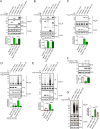
Fig. 4. Nedd4-1(NE) binds VPS33B & VPS16B and ubiquitinates VPS16B.
HEK293T cells were transfected with the indicated constructs. Cell lysates were (AC) subjected to Flag immunoprecipitation (IP), or (D, E, G) incubated in 1% SDS and boiled to dissociate non-covalent protein complexes prior to Flag IP, and the indicated proteins were detected by immunoblotting (Ub: ubiquitin). The ubiquitination signal was normalized to both VPS16B (or VPS33B) IP levels and Nedd4-1(NE) (or Nedd4-1) lysate levels. F Protein levels of Flag-VPS16B were quantified and normalized to cellular actin levels. In (A), Flag blots were imaged at the same time and images were corrected using the same parameters (MW: molecular weight). Constructs labeled “CS” correspond to full-length, catalytically inactive (Cys → Ser) mutants, and the construct labeled E70Q in (G) represents the lymphangiectasia patient mutation. All data are means ± SD of (AE) 3 or (F, G) 4 independent experiments. Statistical significance was determined using (AC) Student’s t-test, or (DG) one-way ANOVA (N.S.: not significant; **: p < 0.01; ***: p < 0.001; ****: p < 0.0001). The markers beside each blot indicate molecular weight (measured in kDa).
Fig. 5
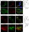
Fig. 5. Nedd4-1(NE) colocalizes with VPS16B in the presence of nutrients.
A, C, D HeLa cells were transfected with the indicated constructs. In (A), cells were incubated for 1 hr in DMEM or HBSS. Cells were fixed, permeabilized, and immunostained with anti-Flag (green) and/or anti-V5 (red) antibodies. Nuclei were stained with DAPI (blue). All scale bars are 10 μm. B, E Quantification of the Pearson correlation coefficient between (B) VPS16B and Nedd4-1(NE) or (E) GFP-LC3 and either Nedd4-1(NE) or Nedd4-1. Each data point represents an individual cell (B) n = 68 DMEM cells, 67 HBSS cells, across 3 independent experiments; (E) n = 86 cells/condition, across 3 independent experiments). Horizontal bars represent the means ± SD. Statistical significance was determined using Student’s t-test (****: p < 0.0001).
Fig. 6
Fig. 6. Nedd4-1(NE) suppresses autophagy.
A HeLa cells were transfected with 3xFlag-Nedd4-1(NE) as indicated. Cells were incubated in either DMEM or HBSS for 4 hrs in the presence or absence of bafilomycin A (100 nM). The markers beside each blot indicate molecular weight (measured in kDa). B Autophagic flux was quantified by measuring endogenous LC3B-II (normalized to total LC3B) stabilization upon bafilomycin A treatment. Data are means ± SD of 5 independent experiments. Statistical significance was determined using Student’s t-test (***: p < 0.001). C Model of the proposed mechanism. When amino acids are abundant, Nedd4-1(NE) localizes to late endosomes (or autophagosomes), resulting in the suppression of autophagy. However, under starvation conditions, IKKβ is activated and phosphorylates Nedd4-1(NE), leading to Nedd4-1(NE) re-localization to the cytosol, thus allowing autolysosomal maturation and autophagy to proceed. Model created in BioRender. Rotin, D. (2025). https://BioRender.com/x91o950.

References

    1. Hershko, A. & Ciechanover, A. The ubiquitin system. Annu. Rev. Biochem.67, 425–479 (1998). - PubMed
    1. Rotin, D. & Prag, G. Physiological functions of the ubiquitin ligases Nedd4-1 and Nedd4-2. Physiol. (Bethesda)39, 18–29 (2024). - PubMed
    1. Plant, P. J., Yeger, H., Staub, O., Howard, P. & Rotin, D. The C2 domain of the ubiquitin protein ligase Nedd4 mediates Ca2+-dependent plasma membrane localization. J. Biol. Chem.272, 32329–32336 (1997). - PubMed
    1. Wiesner, S. et al. Autoinhibition of the HECT-type ubiquitin ligase Smurf2 through its C2 domain. Cell130, 651–662 (2007). - PubMed
    1. Mari, S. et al. Structural and functional framework for the autoinhibition of Nedd4-family ubiquitin ligases. Structure22, 1639–1649 (2014). - PubMed

MeSH terms

Substances

LinkOut - more resources