

Performance of electrochemical aptasensor as antigen test in clinical samples for early diagnosis of leptospirosis
- PMID: 40102450
- PMCID: PMC11920073
- DOI: 10.1038/s41598-025-92685-3
Performance of electrochemical aptasensor as antigen test in clinical samples for early diagnosis of leptospirosis
Abstract
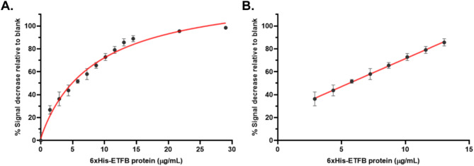
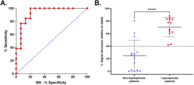
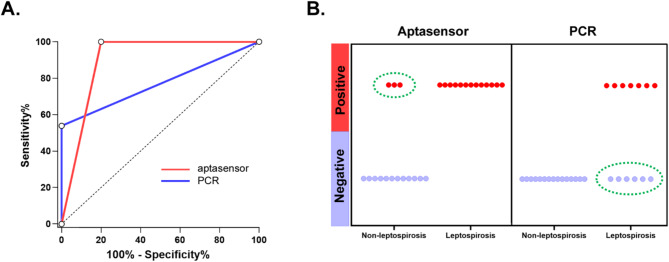
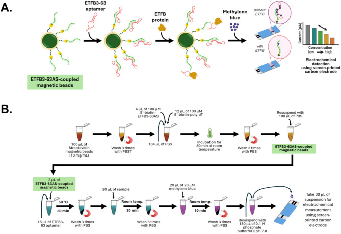
Early diagnosis of leptospirosis is critical for timely treatment and effective disease management. This study evaluated the diagnostic performance of a novel electrochemical aptasensor targeting the electron transfer flavoprotein subunit beta (EtfB) of Leptospira interrogans in clinical samples collected during the acute phase of leptospirosis. The aptasensor assay was tested using plasma samples and compared to the microscopic agglutination test (MAT), the standard reference method. To assess diagnostic performance, aptasensor results were evaluated against leptospirosis status as determined by MAT. Receiver operating characteristic (ROC) analysis identified a 40% decrease in electrochemical signal relative to the blank as the optimal cut-off, yielding an area under the curve (AUC) of 0.93. The assay demonstrated a sensitivity of 100% and a specificity of 80%. For diagnostic concordance, aptasensor results were compared with those obtained from the reference quantitative PCR (qPCR) method. The aptasensor exhibited 100% positive agreement and 57.1% negative agreement with qPCR. Notably, in patients with high MAT titers, the aptasensor outperformed qPCR in detection rates (100% vs. 25%). These findings indicate that the aptasensor assay is a highly reliable and effective antigen-based diagnostic tool for early leptospirosis detection, making it suitable for both low- and high-prevalence settings.
Keywords: Antigen test; Aptamer biosensor; Aptasensor; Diagnostic performance; Early diagnosis; Leptospirosis.
© 2025. The Author(s).
Conflict of interest statement
Declarations. Competing interests: The authors declare no competing interests.
Figures
Similar articles
-
Electrochemical aptasensor detection of electron transfer flavoprotein subunit beta for leptospirosis diagnosis.Analyst. 2023 Sep 25;148(19):4777-4786. doi: 10.1039/d3an01064c. Analyst. 2023. PMID: 37599631
-
Detection of human leptospirosis as a cause of acute fever by capture ELISA using a Leptospira interrogans serovar Copenhageni (M20) derived antigen.BMC Infect Dis. 2013 Sep 20;13:438. doi: 10.1186/1471-2334-13-438. BMC Infect Dis. 2013. PMID: 24053555 Free PMC article.
-
Production of recombinant electron transfer flavoprotein beta subunit protein and its application in a lateral flow assay for early diagnosis of leptospirosis.Med Microbiol Immunol. 2025 Feb 19;214(1):12. doi: 10.1007/s00430-025-00822-6. Med Microbiol Immunol. 2025. PMID: 39969576
-
Nucleic acid and antigen detection tests for leptospirosis.Cochrane Database Syst Rev. 2019 Aug 1;8(8):CD011871. doi: 10.1002/14651858.CD011871.pub2. Cochrane Database Syst Rev. 2019. PMID: 31425612 Free PMC article.
-
Diagnosis of human leptospirosis: systematic review and meta-analysis of the diagnostic accuracy of the Leptospira microscopic agglutination test, PCR targeting Lfb1, and IgM ELISA to Leptospira fainei serovar Hurstbridge.BMC Infect Dis. 2024 Feb 7;24(1):168. doi: 10.1186/s12879-023-08935-0. BMC Infect Dis. 2024. PMID: 38326762 Free PMC article.
References
-
- Daher, E., Zanetta, D. M., Cavalcante, M. B. & Abdulkader, R. C. Risk factors for death and changing patterns in leptospirosis acute renal failure. Am. J. Trop. Med. Hyg.61, 630–634. 10.4269/ajtmh.1999.61.630 (1999). - PubMed
-
- Sehgal, S. C., Murhekar, M. V. & Sugunan, A. P. Outbreak of leptospirosis with pulmonary involvement in North Andaman. Indian J. Med. Res.102, 9–12 (1995). - PubMed
-
- Yersin, C. et al. Pulmonary haemorrhage as a predominant cause of death in leptospirosis in Seychelles. Trans. R Soc. Trop. Med. Hyg.94, 71–76. 10.1016/s0035-9203(00)90445-0 (2000). - PubMed
-
- Plank, R. & Dean, D. Overview of the epidemiology, microbiology, and pathogenesis of Leptospira spp. In humans. Microbes Infect.2, 1265–1276. 10.1016/s1286-4579(00)01280-6 (2000). - PubMed
MeSH terms
Substances
Grants and funding
LinkOut - more resources
Full Text Sources
Miscellaneous
