

Differences in muscle morphology assessed by ultrasound at various muscle regions and their impact on voluntary and involuntary force production
- PMID: 40102558
- PMCID: PMC11920046
- DOI: 10.1038/s41598-025-94158-z
Differences in muscle morphology assessed by ultrasound at various muscle regions and their impact on voluntary and involuntary force production
Abstract
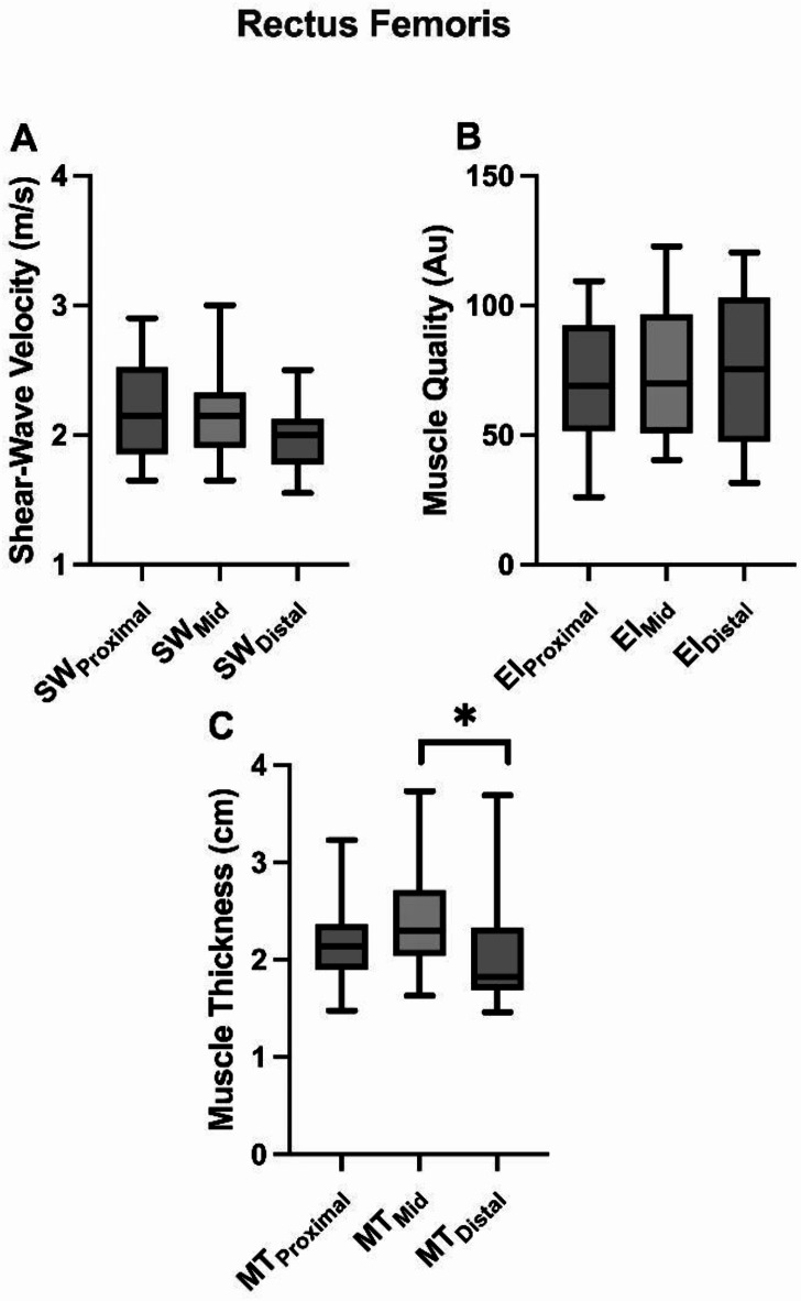
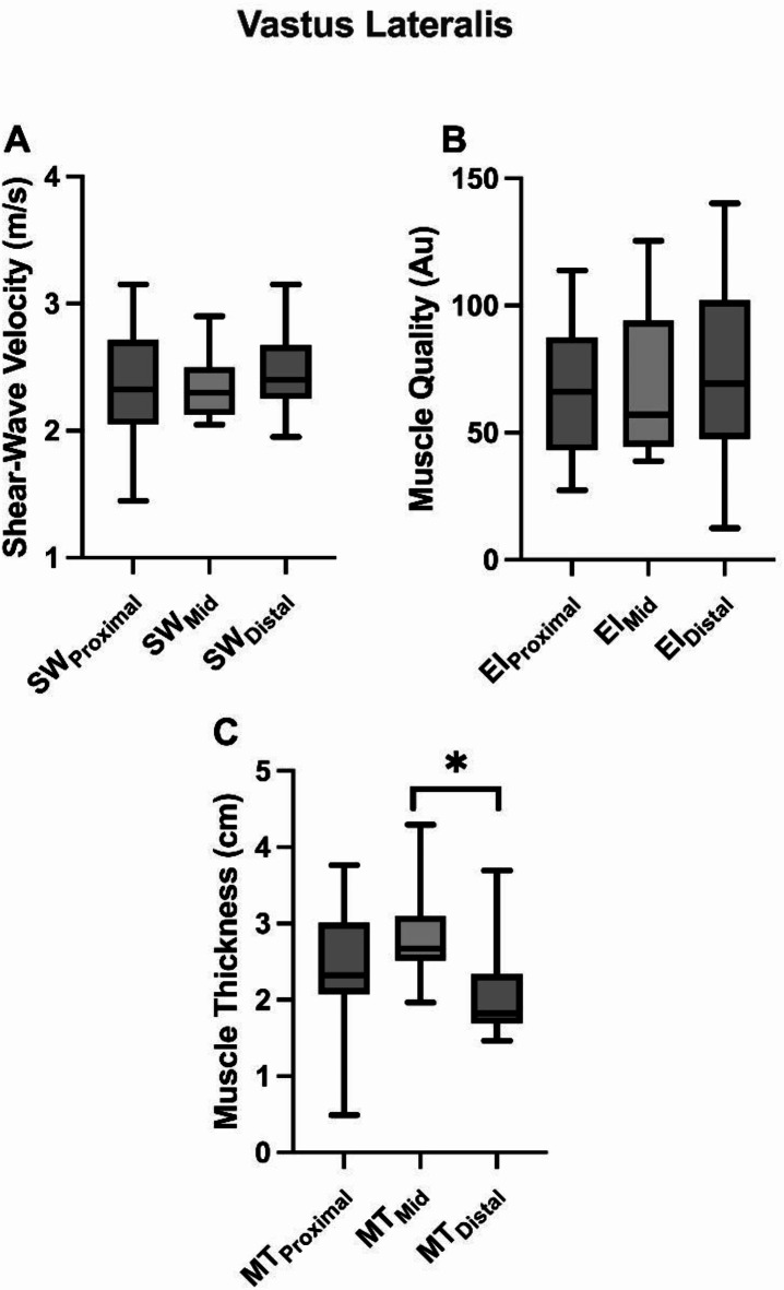
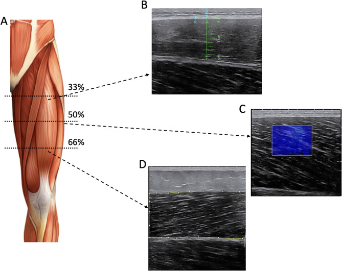
The primary aim of this study was to investigate how measurements from different regions along the rectus femoris (RF) and vastus lateralis (VL) influence muscle morphology, including muscle thickness (MT), muscle stiffness, and muscle quality. An exploratory aim was to examine whether an association exists between voluntary and involuntary force and muscle morphology across the same regions. In one session, participants (n = 13) underwent ultrasound imaging (US), followed by knee extension maximal isometric voluntary contractions and evoked contractions. US recordings (at rest) and testing were conducted while participants were seated at 90º knee flexion (dominant leg) on an isokinetic dynamometer. Muscle morphology was recorded at proximal, medial, and distal regions of RF and VL. During maximum contractions, participants were instructed to exert maximal effort as fast and as forcefully as possible for 5 s, while evoked contractions were performed via femoral nerve stimulation. A one-way repeated measures ANOVA was used for the main aim, while Spearman bivariate correlations were used for the exploratory aim. The primary findings showed that the RF and VL muscles were significantly larger in the medial region (P ≤ 0.023), with no significant differences in muscle quality or stiffness within the same muscle. Additionally, a significant overall relationship was observed between muscle quality and the rate of force development in both muscles (P ≤ 0.037). In conclusion, muscle size varies across the length of the VL and RF muscles, with no changes in muscle quality or stiffness. Furthermore, muscle quality demonstrates a significant association with rate of force development.
Keywords: Muscle quality; Muscle stiffness; Muscle thickness; Ultrasound imaging.
© 2025. The Author(s).
Conflict of interest statement
Declarations. Competing interests: The authors declare no competing interests.
Figures
Similar articles
-
Regional Heterogeneity in Vastus Lateralis Architecture Influences Fascicle Behavior During In Vivo Contractions.Scand J Med Sci Sports. 2025 Jul;35(7):e70103. doi: 10.1111/sms.70103. Scand J Med Sci Sports. 2025. PMID: 40631593 Free PMC article.
-
Dose-Response Relationships of Resistance Training in Healthy Old Adults: A Systematic Review and Meta-Analysis.Sports Med. 2015 Dec;45(12):1693-720. doi: 10.1007/s40279-015-0385-9. Sports Med. 2015. PMID: 26420238 Free PMC article.
-
Physical exercise training interventions for children and young adults during and after treatment for childhood cancer.Cochrane Database Syst Rev. 2013 Apr 30;(4):CD008796. doi: 10.1002/14651858.CD008796.pub2. Cochrane Database Syst Rev. 2013. Update in: Cochrane Database Syst Rev. 2016 Mar 31;3:CD008796. doi: 10.1002/14651858.CD008796.pub3. PMID: 23633361 Updated.
-
Physical exercise training interventions for children and young adults during and after treatment for childhood cancer.Cochrane Database Syst Rev. 2016 Mar 31;3(3):CD008796. doi: 10.1002/14651858.CD008796.pub3. Cochrane Database Syst Rev. 2016. PMID: 27030386 Free PMC article.
-
Skeletal Muscle Compliance and Echogenicity in Resistance-Trained and Nontrained Women.J Strength Cond Res. 2024 Apr 1;38(4):671-680. doi: 10.1519/JSC.0000000000004669. Epub 2023 Oct 16. J Strength Cond Res. 2024. PMID: 38513175 Free PMC article.
References
-
- Evangelidis, P. E., Massey, G. J., Pain, M. T. G. & Folland, J. P. Strength and size relationships of the quadriceps and hamstrings with special reference to reciprocal muscle balance. Eur. J. Appl. Physiol.116, 593–600 (2016). - PubMed
-
- Ando, R. & Suzuki, Y. Positive relationship between passive muscle stiffness and rapid force production. Hum. Mov. Sci.66, 285–291 (2019). - PubMed
MeSH terms
Grants and funding
LinkOut - more resources
Full Text Sources
