. 2025 Mar 18;15(1):9280.
doi: 10.1038/s41598-025-94414-2.

Centrobin serves as a safeguard to guide timely centriole maturation during the cell cycle

Affiliations

Centrobin serves as a safeguard to guide timely centriole maturation during the cell cycle

Dohyong Lee et al. Sci Rep. .

Abstract

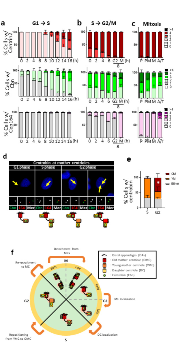
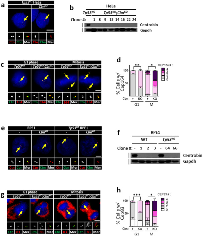
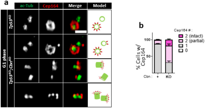
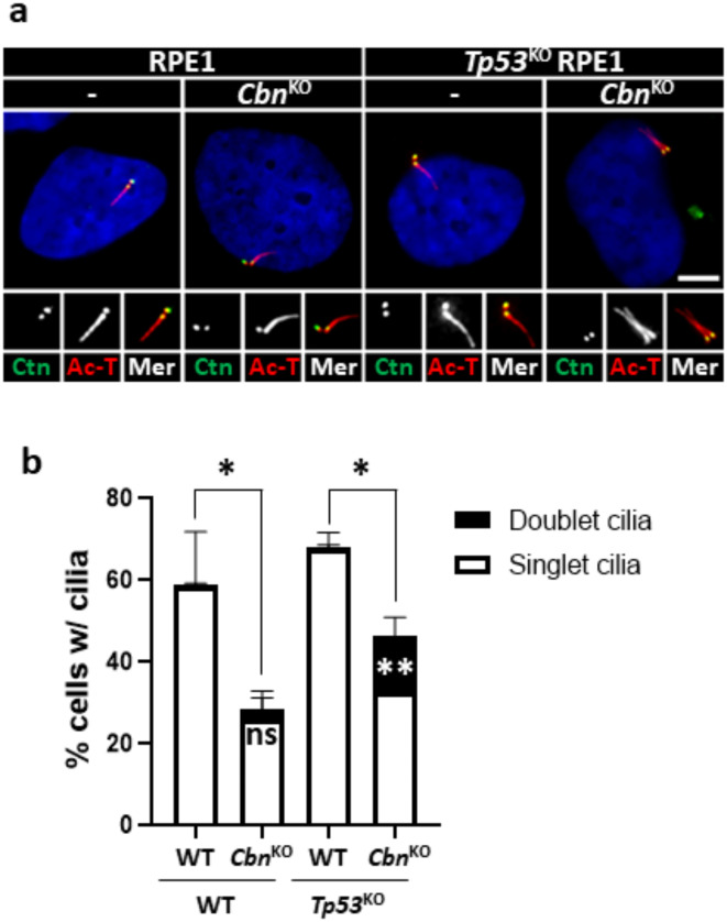
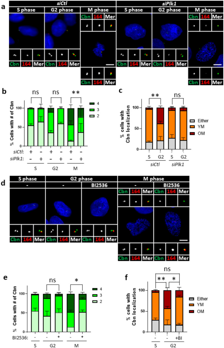
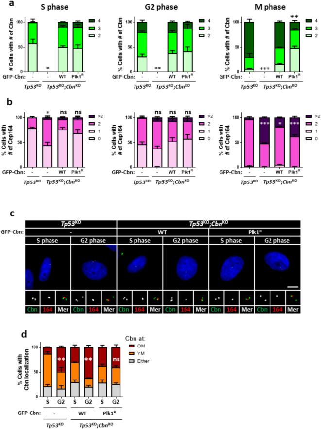
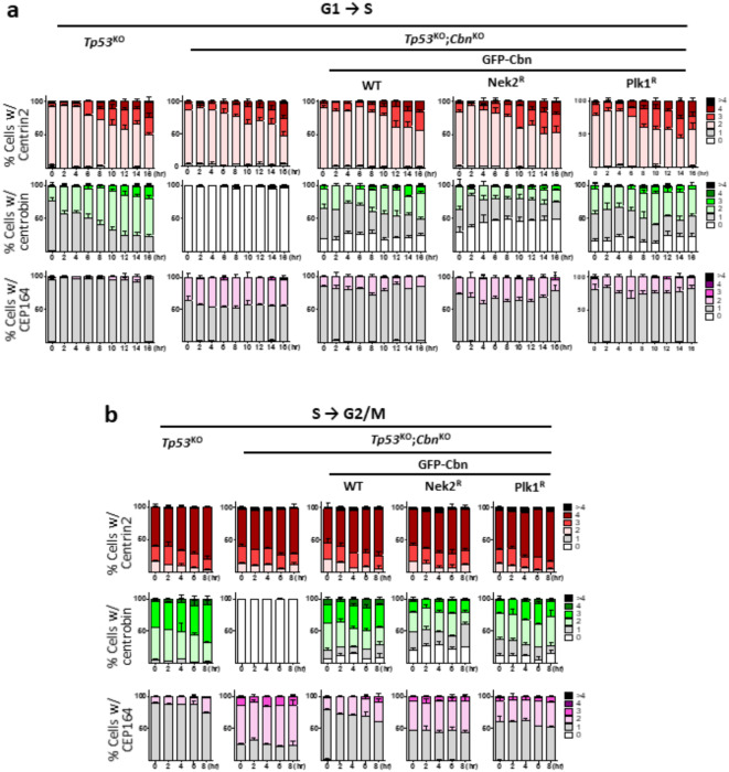
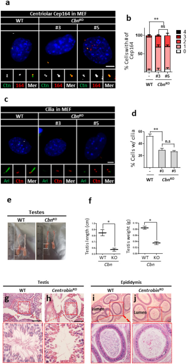
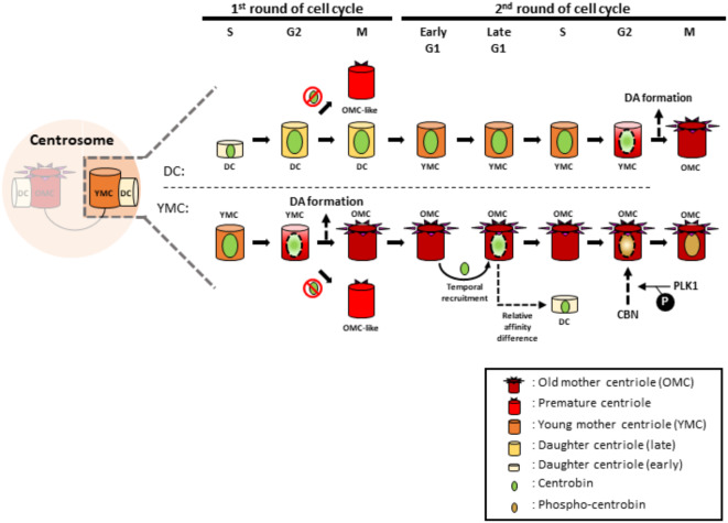
Centrioles assemble and segregate in link to the cell cycle. Daughter centrioles assemble at S phase, and become young mother centrioles after M phase. Since distal appendages (DAs) are installed to young mother centrioles at the second G2/M transition phase, it takes one and a half cell cycle for a daughter centriole to fully mature into an old mother centriole. Here, we investigated specific roles of centrobin on centriole maturation by tracing its centriole localization throughout the cell cycle. Centrobin instantly places at the nascent daughter centrioles during the S phase, maintains its localization through subsequent cell cycle as these daughter centrioles mature into young mother centrioles, and detaches from the young mother centriole during the G2 phase, prior to DA installation. Centrobin KO cells exhibit two DA-installed centrioles, due to premature DA installation in daughter centrioles, and can produce doublet cilia from two DA-installed basal bodies. We also present evidence that direct phosphorylation of Plk1 is crucial for centrobin attachment to centrioles during G2 and M phases. Finally, premature DA installation was also observed in centrobin KO mice. Our results collectively demonstrate that centrobin serves as a safeguard to guide timely centriole maturation during the cell cycle.

Keywords: Cell cycle; Centriole; Centriole maturation; Centrobin; Distal appendage; Plk1; Spermatogenesis.


Conflict of interest statement

Declarations. Competing interests: The authors declare no competing interests.

Figures

Fig. 1
Fig. 1
Centriolar localization of centrobin during the cell cycle (a) HeLa cells at G1 phase were prepared with the mitotic shake-off method, and synchronously cultured for up to 16 h to reach to the S phase. (b) HeLa cells at S phase were prepared with the double thymidine block and release method, and cultured for up to 8 h to reach to the M phase. (c) HeLa cells at M phase were enriched with the thymidine-RO3306 release method. Mitotic stages of the individual cells were determined with the DAPI staining. (ac) The cells at indicated time points were coimmunostained with centrin2, centrobin and Cep164 antibodies. Representative images of the staining patterns are shown in Supplementary Fig. 1. The number of centriole signals per cell was counted. (d) The cells were arrested at G1/S transition phase with the double thymidine block method, and synchronously released for 2, 6 and 8 h to reach to S, G2 and G1 phases, respectively. The cells were coimmunostained with centrobin (green) and Cep164 (red) antibodies. Scale bar, 10 μm. (e) The number of cells with centrobin signals at young and old mother centrioles was counted. Statistical significance was determined using paired t-test. *, P < 0.05. (a-c, e) More than 90 cells per group were counted in 3 independent experiments. (f) Summary of the centriolar localization of centrobin during the cell cycle.
Fig. 2
Fig. 2
Precocious installation of DAs in the centrobinKO centrioles (a) The Tp53KO;centrobinKO HeLa cells were arrested at G1 and M phases, and coimmunostained with centrin2 (red) and centrobin (green) antibodies. (b) Several lines of the Tp53KO;centrobinKO HeLa cells were subjected to immunoblot analyses with centrobin and Gapdh antibodies. (c) The Tp53KO;centrobinKO HeLa cells at G1 and M phases were coimmunostained with the centrin2 (green) and Cep164 (red) antibodies. (d) The number of centriolar Cep164 signals per cell was counted in the Tp53KO;centrobinKO HeLa cells. (e) The centrobinKO and TP53KO;centrobinKO RPE1 cells were coimmunostained with centrin2 (red) and centrobin (green) antibodies. (f) Several lines of the centrobinKO and Tp53KO;centrobinKO RPE1 cells were subjected to immunoblot analyses with centrobin and Gapdh antibodies. (g) The centrobinKO RPE1 cells at G1 and M phases were coimmunostained with centrin2 (green) and Cep83 (red) antibodies. (h) The number of centriolar Cep83 signals per cell was counted in the centrobinKO RPE1 cells. (a, c,e, g) Scale bars, 10 μm. (d, h) More than 90 cells per group were counted in 3 independent experiments. Statistical significance was determined using paired t-test. *, P < 0.05.
Fig. 3
Fig. 3
Observation of DAs in centrobinKO cells with super-resolution microscopy (a) The Tp53KO;centrobinKO HeLa cells were coimmunostained with acetylated α-tubulin (green) and Cep164 (red) antibodies, and observed with super-resolution microscopy. Scale bar, 1 μm. (b) The number of centriolar Cep164 signals per cell was counted. More than 30 cells per group were counted in 3 independent experiments.
Fig. 4
Fig. 4
Cilia formation in centrobinKO RPE1 cells (a) The centrobinKO and Tp53KO;centrobinKO RPE1 cells were cultured in a serum-deprived medium for 48 h, and coimmunostained with centrin2 (green) and acetylated α-tubulin (red) antibodies. Scale bar, 10 μm. (b) The number of cells with singlet and doublet cilia were counted. More than 90 cells per group were counted in 3 independent experiments. Statistical significance was determined using unpaired t-test. *, P < 0.05; **, P < 0.01.
Fig. 5
Fig. 5
Plk1 regulation of centrobin localization at mitosis (a) Plk1-depleted HeLa cells were arrested at S, G2, and M phases, and coimmunostained with centrobin (green) and Cep164 (red) antibodies. (b) The number of centrobin signals per cell was counted at indicated cell cycle stages. (c) The number of cells with centrobin signals at young and old mother centrioles was quantified. (d) HeLa cells at S, G2, and M phases were treated with BI2536 for 6 h, and coimmunostained with centrobin (green) and Cep164 (red) antibodies. (e) The number of centrobin signals were counted in cells at indicated cell cycle stages. (f) The number of cells with centrobin signals at young and old mother centrioles was quantified. (a, d) Scale bars, 10 μm. (b, c,e, f) More than 90 cells per group were counted in 3 independent experiments. Statistical significance was determined using paired t-test. *, P < 0.05; **, P < 0.01.
Fig. 6
Fig. 6
Plk1 phosphorylation of centrobin for its localization at mitosis Tp53KO;centrobinKO HeLa cells were stably rescued with the wild type (GFP-centrobin) and Plk1-phospho-resistant mutant centrobin (GFP-CbnPlk1R). (a, b) The cells were arrested at S, G2, and M phases and coimmunostained with centrobin and Cep164 antibodies. The number of centrobin (a) and Cep164 (b) signals per cell was counted. (c) The cells were arrested at G1/S transition phase with the double thymidine block method, and synchronously released for 2 and 6 h to reach to S and G2 phases, respectively. The cells were coimmunostained with centrobin (green) and Cep164 (red). Scale bar, 10 μm. (d) The number of cells with centrobin signals at young and old mother centrioles was counted. (a, b, d) More than 90 cells per group were counted in 3 independent experiments. Statistical significance was determined using paired t-test. *, P < 0.05; **, P < 0.01.
Fig. 7
Fig. 7
Nek2 phosphorylation of centrobin for its localization at interphase Tp53KO;centrobinKO HeLa cells were stably rescued with the wild type (GFP- Cbn), Nek2-phospho-resistant mutant centrobin (GFP-CbnNekR), and Plk1-phospho-resistant mutant centrobin (GFP-CbnPlkR). (a) Mitotic cells were isolated using the shake-off method and re-plated to allow synchronous entry into the G1 phase. (b) The cells were arrested at G1/S transition phase with the double thymidine block method and synchronously released to S phase. (a, b) The cells were collected at 2-hour intervals and coimmunostained with centrin2, centrobin, and Cep164 antibodies. The numbers of centrin2, centrobin, and Cep164 signals per cell were counted at each time point. More than 90 cells per group were counted in 3 independent experiments.
Fig. 8
Fig. 8
Analysis of centrobin KO mice (a) Centrobin KO MEF lines were coimmunostained with centrin2 (green) and Cep164 (red) antibodies. (b) The number of centriolar Cep164 signals per cell was counted. (c) Centrobin KO MEF lines were coimmunostained with Arl13b (green) and centrin2 (red) antibodies. (d) The number of cells with cilia was counted. (e, f) The sizes of testes from the wild-type and centrobin KO mice were measured. At least three mice per group were used. (gj) Testes and epididymis of the wild type and centrobin KO mice were subjected to H&E staining analyses. Scale bars, 100 μm. (a, c) Scale bars, 10 μm. (b, d) More than 90 cells per group were counted in 3 independent experiments. Statistical significance was determined using paired t-test. *, P < 0.05; **, P < 0.01.
Fig. 9
Fig. 9
Summary Once centrobin is placed at nascent daughter centrioles, it lasts at the centrioles for one and a half cell cycle stages, and released before the time when DAs are about to be installed. Indeed, DAs are prematurely installed at daughter centrioles in centrobin KO cells. Centriole localization of centrobin is regulated by Plk1.

Similar articles

References

    1. Kong, D. & Loncarek, J. Analyzing centrioles and cilia by expansion microscopy. Methods Mol. Biol.2329, 249–263. 10.1007/978-1-0716-1538-6_18 (2021). - PMC - PubMed
    1. Fu, J. et al. Conserved molecular interactions in centriole-to-centrosome conversion. Nat. Cell. Biol.18, 87–99. 10.1038/ncb3274 (2016). - PMC - PubMed
    1. Fong, C. S., Ozaki, K. & Tsou, M. B. PPP1R35 ensures centriole homeostasis by promoting centriole-to-centrosome conversion. Mol. Biol. Cell.29, 2801–2808. 10.1091/mbc.E18-08-0525 (2018). - PMC - PubMed
    1. Atorino, E. S., Hata, S., Funaya, C., Neuner, A. & Schiebel, E. CEP44 ensures the formation of Bona Fide centriole wall, a requirement for the centriole-to-centrosome conversion. Nat. Commun.11, 903. 10.1038/s41467-020-14767-2 (2020). - PMC - PubMed
    1. Sullenberger, C., Vasquez-Limeta, A., Kong, D. & Loncarek, J. With age comes maturity: biochemical and structural transformation of a human centriole in the making. Cells9, 1429. 10.3390/cells9061429 (2020). - PMC - PubMed

Substances

LinkOut - more resources