. 2025 Mar;13(6):e70237.
doi: 10.14814/phy2.70237.

Corticomuscular and intermuscular coherence during evidence accumulation in sensorimotor decision-making

Affiliations

Corticomuscular and intermuscular coherence during evidence accumulation in sensorimotor decision-making

Yvonne F Visser et al. Physiol Rep. 2025 Mar.

Abstract

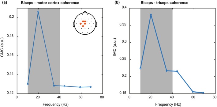
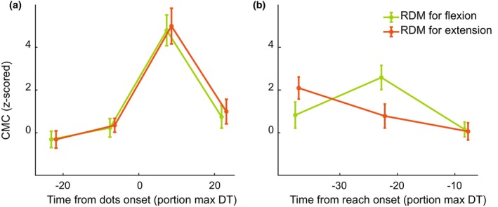
Evidence accumulation processes during decision-making are thought to continuously feed into the motor system, preparing multiple competing motor plans, of which one is executed when the evidence is complete. Previously, the state of this accumulation process has been studied by reading out the preparatory state of the motor system with evoked responses, once per trial. In this study, we aim to continuously track the sensorimotor decision during the trial using corticomuscular (CMC) and intermuscular coherence (IMC). We recorded EEG and EMG of healthy young adults (n = 34) who viewed random dot motion stimuli, with varying strengths across trials, and indicated their perceived motion direction by reaching towards one of two targets, requiring either flexion or extension of the elbow. Coherence was computed in the beta band. After stimulus presentation, both CMC and IMC show an initial phasic pattern, which is followed by sustained coherence patterns at a level that depends on stimulus strength for CMC. Prior to reach onset, the CMC for different stimulus strengths had a tendency to settle at similar levels. This tendency tentatively marks a stimulus-independent decision bound. We conclude that CMC, and to a lesser extent IMC, track the evidence accumulation process on a single trial.

Keywords: corticomuscular coherence; decision‐making; evidence accumulation; intermuscular coherence; motor control.


Conflict of interest statement

The authors declare no conflicts of interest.

Figures

FIGURE 1
FIGURE 1
Experimental design. The middle row shows a schematic of a single trial. Participants had to keep their hand cursor (blue dot) on the central fixation cross (red). Next, the fixation cross was replaced by an RDM stimulus (the number of dots in this figure are schematic and do not reflect actual stimulus parameters). Participants had to reach to the target that matched the inferred direction of the stimulus. In this case, a reach towards the extension target is plotted, requiring triceps activation and relaxation of the biceps. Participants received feedback about their choice after the reach (target turning green or red), and the robotic manipulandum brought the hand back to the central location (white dashed line). The trajectory of the cursor was never visible to the participant and only plotted here, in red, for clarity. The upper row shows windowing for the stimulus‐aligned coherence analysis. Windows are expressed as a percentage of the longest decision time (DT) per participant. The first two windows occur before stimulus onset and the last two windows during the RDM presentation. In the lower row the windows are aligned to reach onset. All windows occur during the RDM presentation, before reach onset. Arrival in the chosen target is indicated with a vertical dashed line around 20% of normalized decision time.
FIGURE 2
FIGURE 2
Behavioral performance, directional EMG activity, and readiness potential. (a) Grand average (n = 34) of accuracy of choices per stimulus strength. (b) Grand average of decision time per stimulus strength. (c) Grand average EMG response for the biceps when reaching for the flexion versus extension target. (d) Grand average EMG response for the triceps when reaching for the flexion versus the extension target. (e) Grand average preparatory activity (i.e., readiness potential) over the Cz electrode prior to reach onset. Shaded areas in all panels indicate S.E.M.
FIGURE 3
FIGURE 3
Power spectra of CMC and IMC. (a) Frequency spectrum of the corticomuscular coherence between biceps and the motor cortex near reach onset. (b) Frequency spectrum of the intermuscular coherence between biceps and triceps near reach onset. Gray areas indicate the part of the frequency spectrum that was included in the rest of the analysis for both panels. Note that the high‐pass filter applied to the EMG data prevents us from considering frequencies lower than 10 Hz.
FIGURE 4
FIGURE 4
CMC development over time. Corticomuscular coherence (solid) and intermuscular coherence (dashed) relative to stimulus and reach onset (vertical dashed lines). All stimulus strengths were averaged together. Error bars represent S.E.M., and markers on the x‐axis are staggered for plotting purposes only.
FIGURE 5
FIGURE 5
Corticomuscular coherence of the biceps as a function of time relative to stimulus onset (left column) and relative to reach onset (right column). The top row represents trials in which the stimulus direction was towards the lower left target (flexion) while the bottom row represents those in which the stimulus direction was towards the upper right target (extension). Trials with strong stimulus strength (25.6% and 51.2%) are averaged together and plotted in red, while trials with weak stimulus strength (3.2%, 6.4%, and 12.8%) are plotted in blue. Error bars represent S.E.M., and markers on the x‐axis are staggered for plotting purposes only.
FIGURE 6
FIGURE 6
Intermuscular coherence as a function of time relative to stimulus onset (left column) and relative to reach onset (right column). The top row represents trials in which the stimulus direction was towards the lower left target (flexion) while the bottom row represents those in which the stimulus direction was towards the upper right target (extension). Trials with strong stimulus strength (25.6% and 51.2%) are averaged together and plotted in red, while trials with weak stimulus strength (3.2%, 6.4%, and 12.8%) are plotted in blue. Error bars represent S.E.M., and markers on the x‐axis are staggered for plotting purposes only.
FIGURE 7
FIGURE 7
Corticomuscular coherence of the biceps for different stimulus directions. CMC was aligned to (a) stimulus onset or (b) reach onset. Trials in which the stimulus was in the direction of the flexion target were averaged together and plotted in green, while the average of all trials in which the stimulus was in the direction of the extension target is plotted in orange. Error bars represent S.E.M., and markers on the x‐axis are staggered for plotting purposes only. The same results for triceps CMC, as well as IMC can be found in Figure S1.

Similar articles

References

    1. Baker, M. R. , & Baker, S. N. (2003). The effect of diazepam on motor cortical oscillations and corticomuscular coherence studied in man. Journal of Physiology, 546(Pt 3), 931–942. 10.1113/jphysiol.2002.029553 - DOI - PMC - PubMed
    1. Baker, S. N. (2007). Oscillatory interactions between sensorimotor cortex and the periphery. Current Opinion in Neurobiology, 17(6), 649–655. 10.1016/j.conb.2008.01.007 - DOI - PMC - PubMed
    1. Baker, S. N. , Olivier, E. , & Lemon, R. N. (1997). Coherent oscillations in monkey motor cortex and hand muscle EMG show task‐dependent modulation. Journal of Physiology, 501(Pt 1), 225–241. 10.1111/j.1469-7793.1997.225bo.x - DOI - PMC - PubMed
    1. Bayraktaroglu, Z. , von Carlowitz‐Ghori, K. , Curio, G. , & Nikulin, V. V. (2013). It is not all about phase: Amplitude dynamics in corticomuscular interactions. NeuroImage, 64, 496–504. 10.1016/j.neuroimage.2012.08.069 - DOI - PubMed
    1. Boonstra, T. W. , & Breakspear, M. (2012). Neural mechanisms of intermuscular coherence: Implications for the rectification of surface electromyography. Journal of Neurophysiology, 107(3), 796–807. 10.1152/jn.00066.2011 - DOI - PubMed

LinkOut - more resources