. 2025 Mar 18;23(1):164.
doi: 10.1186/s12916-025-03987-2.

Cellular sentinels: empowering survival and immune defense in hematopoietic stem cell transplantation through mesenchymal stem cells and T lymphocytes

Affiliations

Cellular sentinels: empowering survival and immune defense in hematopoietic stem cell transplantation through mesenchymal stem cells and T lymphocytes

Tzong-Shyuan Tai et al. BMC Med. .

Abstract

Background: Hematopoietic stem cell transplantation (HSCT) is a critical treatment for hematologic disorders such as leukemia, lymphoma, and specific immune deficiencies. Despite its efficacy, challenges such as engraftment failure and delayed neutrophil regeneration remain significant barriers. These complications lead to prolonged cytopenia, increased risks of infections and other complications, and elevated morbidity and mortality rates. While mesenchymal stem cells (MSCs) are known to play essential roles in supporting hematopoiesis, the precise mechanisms and interactions between MSCs and other cellular components in HSCT require further investigation.

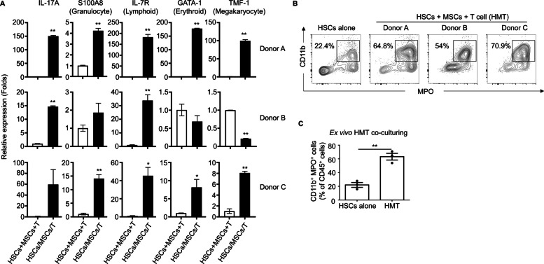
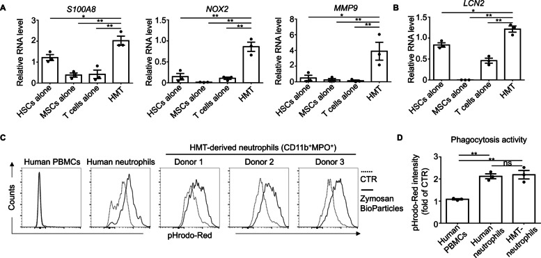
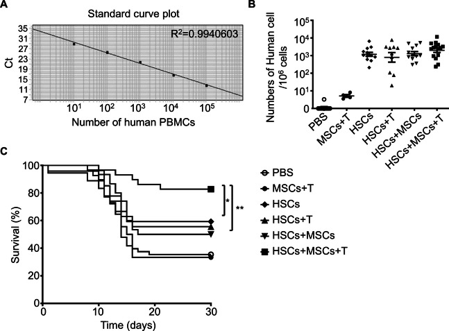
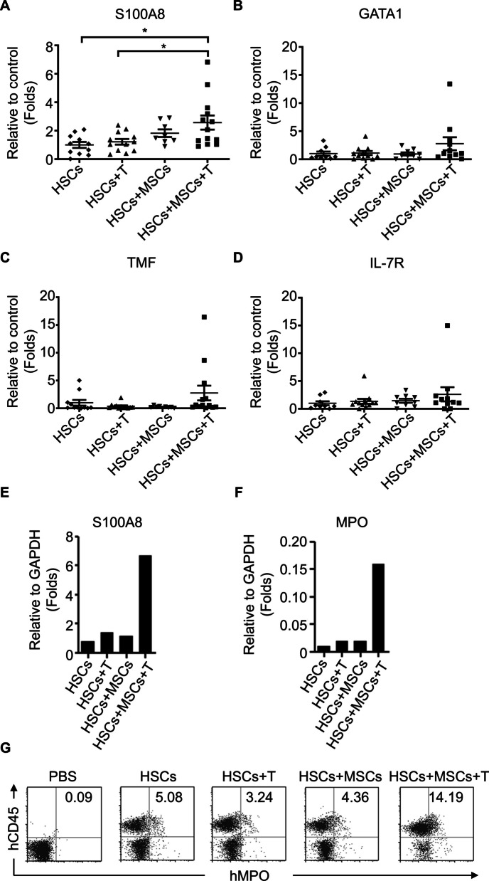
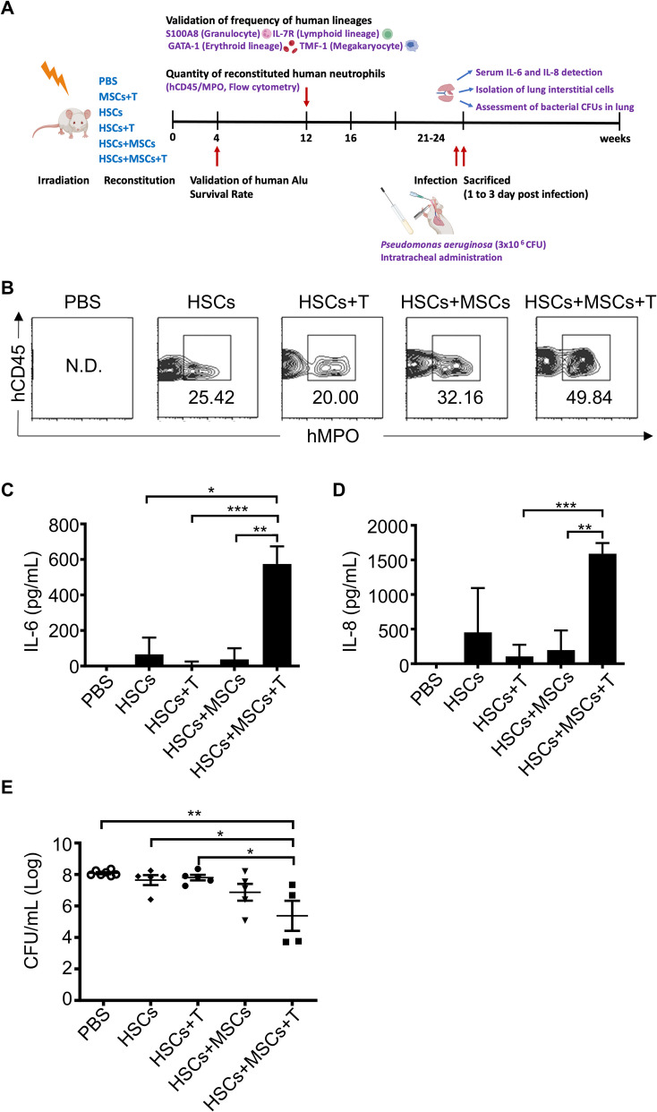
Methods: To address these challenges, we explored the combined infusion of allotype-cord blood hematopoietic stem cells (HSCs) and activated T cells from the same donor along with third-party MSCs. The study assessed the effects of this triple-cell therapy on neutrophil differentiation and function ex vivo and in vivo. Using a respiratory infection model, we evaluated the accumulation of human neutrophils, cytokine secretion (IL-6 and IL-8), bacterial clearance, and overall survival compared to control groups.

Results: The triple-cell therapy demonstrated a significant improvement in the differentiation of human HSCs into neutrophils both in ex vivo and in vivo. In the respiratory infection model, this approach resulted in enhanced accumulation of human neutrophils, increased secretion of IL-6 and IL-8, superior bacterial clearance, and reduced mortality rates compared to the control group. These findings highlight the synergistic interplay between allo-HSCs, MSCs, and activated T cells in promoting neutrophil production and function.

Conclusions: Our study presents a novel therapeutic strategy combining allo-HSCs, activated T cells, and third-party MSCs to enhance neutrophil production and functionality post-transplantation. This approach not only accelerates neutrophil regeneration but also improves resistance to infections, offering a promising avenue to overcome engraftment challenges in HSCT.

Keywords: Activated T cells; Engraftment enhancement; Hematopoietic stem cell transplantation (HSCT); Mesenchymal stem cells (MSCs); Neutrophil regeneration.


Conflict of interest statement

Declarations. Ethics approval and consent to participate: The study was performed in accordance with the Declaration of Helsinki and approved by the institutional review board of Taoyuan General Hospital, Ministry of Health and Welfare, Taiwan (TYGH100040, TYGH109026), and the Research Ethics Committee of National Health Research Institutes (EC1000802, EC1001101-R1, EC1030604-E, EC1111003-W, EC1000802, EC1030604-E, EC1111215-W, and EC1100308-E). All written informed consents were obtained from participants of this study. Consent for publication: All authors consent to publication. Competing interests: The authors declare no competing interests.

Figures

Fig. 1
Fig. 1
Illustrates the guidance of human hematopoietic stem cell (HSC) differentiation toward specialized immune lineages with human bone marrow-derived mesenchymal stem cells (BM-MSCs) and CD3+ T cells. A Human CD34+HSCs (1 × 105) were isolated from three donors and cultured alone or co-cultured with allogenic MSCs (2 × 104) and activated stem cell donor-matched T cells (1 × 10.5) (HSCs/MSCs/T) for 14 days ex vivo. After that, cells were harvested, and their total RNA was extracted to examine the expression of specific genes using quantitative real-time PCR. RNA from HSCs alone, MSCs alone, and T cell alone was evenly mixed in equal amounts as the control group sample, HSCs + MSCs + T. The expression levels of representative genes, including S100A8 (Granulocyte lineage), IL-7R (Lymphoid lineage), GATA-1 (Erythroid lineage), and TMF-1 (Megakaryocyte lineage), are depicted as folds of the control level. Suspensive HSC cells alone and the combination of (HSCs + MSCs + T) from three various HSC donors were harvested at 14 days, and the CD11b⁺ MPO⁺ population was identified using flow cytometry (B). The statistical analysis of ex vivo differentiation from three different donors is summarized in C. Data were expressed as mean ± S.E.M., and significant differences between groups were indicated (*p < 0.05, **p < 0.01; two-sided unpaired t test, ns, not significant)
Fig. 2
Fig. 2
Co-culture of human CD34+ HSCs with human MSCs and autologous T cells enhances granulocytic lineage development and phagocytic function. Human CD34+ HSCs were cultured alone or co-cultured with BM-MSCs and autologous CD3+ T cells at defined ratios (HSCs: 1 × 10⁵ cells, T cells: 1 × 10⁵ cells, MSCs: 2 × 10⁴ cells) with 4 × 10³ CD3/CD28 T-cell activator beads for 14 daysin vitro. Total RNA was extracted from cultured cells and analyzed by real-time PCR to quantify mRNA expression of neutrophil-associated markers, including S100A8, NOX2, MMP9, and LCN2(panels A, B). Phagocytic function was assessed by co-culturing suspension cells with 100 µg/ml pHrodo-red bioparticles for 2 h, with subsequent analysis of the CD11b⁺ MPO⁺ cell population (panel C). Phagocytic activity was determined across human peripheral blood mononuclear cells (PBMCs), freshly isolated neutrophils, and hematopoietic microenvironment (HMT)-derived CD11b⁺ MPO⁺ cells. Data are presented as means ± standard error of the mean (S.E.M.) from three independent experiments. Statistical significance was assessed using a two-sided unpaired t test (*p < 0.05, **p < 0.01, ns = not significant)
Fig. 3
Fig. 3
Co-transplantation of human HSCs with human MSCs and T cells enhanced survival of irradiated NOD/SCID mice. A A standard curve for the calculation of human cell numbers. Human peripheral blood mononuclear cells (PBMCs) were tenfold serially diluted (106, 105, 104, 103, 102, 101, 100 cells) and mixed with mouse splenocytes to a total cell number of 1 × 106. Each mixture was subjected to genomic DNA extraction for the real-time PCR analysis to quantify human Alu repeat sequence and mouse albumin. A standard curve is presented as a semi-log regression line plot of mean Ct values versus the logarithm of input human PBMC numbers. B Measurement of human-derived cells in irradiated NOD/SCID mice after transplantation. NOD/SCID mice (4-week-old) were given 2000 mGy irradiation and then received an intravenous infusion of PBS or various combinations of human HSCs (1 × 105), MSCs (5 × 105), and autologous T cells (1 × 10.5) as indicated (PBS: ○, n = 10; MSCs + T: ●, n = 4; HSCs: ◆, n = 15; HSCs + T: ▲, n = 14; HSCs + MSCs: ▼, n = 14; HSCs + MSCs + T: ■, n = 23). After 4 weeks of transplantation, the peripheral blood of recipient mice was analyzed for the content of human Alu repeat sequence by real-time PCR, and the number of human-derived cells was calculated from the Ct value using the standard curve mentioned above. Data were expressed as mean ± S.E.M., and significant differences between groups were indicated (*p < 0.05, **p < 0.01; one-way ANOVA). C Survival curves of irradiated NOD/SCID mice after transplantation. After cell transplantation, the survival of recipient mice was followed up for 30 days. Survival curves were analyzed for the differences between groups using the Log-rank test, and significant differences were indicated (*p < 0.05, **p < 0.01)
Fig. 4
Fig. 4
Co-transplantation of human HSCs with T cells and MSCs demonstrated granulocytic lineage development. Leukocytes were isolated from the peripheral blood of humanized NOD/SCID mice that were transplanted with various combinations of human cord blood HSCs (1 × 105), MSCs (5 × 105), and autologous T cells (1 × 105) as indicated (HSCs: ◆, n = 11; HSCs + T: ▲, n = 12; HSCs + MSCs: ▼, n = 8; HSCs + MSCs + T: ■, n = 13). Total RNA was purified from the isolated leukocytes and analyzed by the real-time PCR to assess the mRNA levels of representative cell-lineage markers, including A S100A8 (Granulocyte), B GATA1 (Erythrocyte), C TMF1 (Megakaryocyte), and D IL7R (Lymphocyte). Additionally, bone marrow cells were harvested for evaluating the gene expression of human S100A8 (E) and MPO (F) by real-time PCR. G These bone marrow cells were also subjected to flow cytometry analysis to determine the expression of hCD45 and hMPO
Fig. 5
Fig. 5
Enhanced anti-Pseudomonal defense via neutrophil reconstitution by co-transplantation of MSCs and T cells in recipients of human HSCs. A Experimental timeline. NOD/SCID mice were given sub-lethal irradiation and then transplanted with PBS (as negative control) or different combinations of human HSCs, MSCs, and T cells. At 4 and 12 weeks after transplantation, mice were assessed for human Alu signal and quantified for reconstituted human HSCs, respectively. Subsequently, after 21 weeks of HSC reconstitution, mice were anesthetized with freshly prepared 2% avertin (0.019 mL/g) via intraperitoneal injection, followed by infection with a half-lethal dose (LD50) (~ 3 × 10 6 CFU, colony formation unit) of Pseudomonas aeruginosa for 1 day to induce acute pulmonary infection. Lung tissues were collected, and lung interstitial cells (mCD146+cells) were isolated and gated with hCD45 to determine the frequency of human CD45+MPO.+ cells (B). The expression levels of human IL-6 (C) and human IL-8 (D) in the sera collected from infected mice were examined via ELISA. E The lung tissues of infected groups were collected, disrupted with a tissue chopper, and plated on LB agar plates to calculate bacterial load (CFU). Data were expressed as mean ± S.E.M., and significant differences between groups were indicated (*p < 0.05, **p < 0.01; two-sided unpaired t-test, ns, not significant)

Similar articles

References

    1. Orkin SH, Zon LI. Hematopoiesis: an evolving paradigm for stem cell biology. Cell. 2008;132(4):631–44. - PMC - PubMed
    1. Crane GM, Jeffery E, Morrison SJ. Adult haematopoietic stem cell niches. Nat Rev Immunol. 2017;17(9):573–90. - PubMed
    1. Copelan EA. Hematopoietic stem-cell transplantation. N Engl J Med. 2006;354(17):1813–26. - PubMed
    1. Adams GB, Martin RP, Alley IR, Chabner KT, Cohen KS, Calvi LM, Kronenberg HM, Scadden DT. Therapeutic targeting of a stem cell niche. Nat Biotechnol. 2007;25(2):238–43. - PubMed
    1. Calvi LM, Adams GB, Weibrecht KW, Weber JM, Olson DP, Knight MC, Martin RP, Schipani E, Divieti P, Bringhurst FR, et al. Osteoblastic cells regulate the haematopoietic stem cell niche. Nature. 2003;425(6960):841–6. - PubMed

MeSH terms

LinkOut - more resources