Comparative Study
. 2025 Mar 19;21(1):181.
doi: 10.1186/s12917-025-04648-5.

Comparative analysis of iridocorneal angle in cats and dogs using ultrasound biomicroscopy: implications for glaucoma prevalence

Affiliations
Comparative Study

Comparative analysis of iridocorneal angle in cats and dogs using ultrasound biomicroscopy: implications for glaucoma prevalence

Donghee Kim et al. BMC Vet Res. .

Abstract

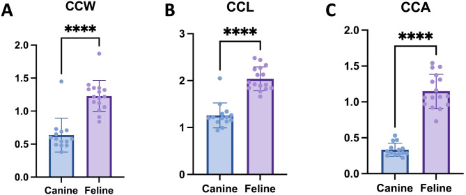
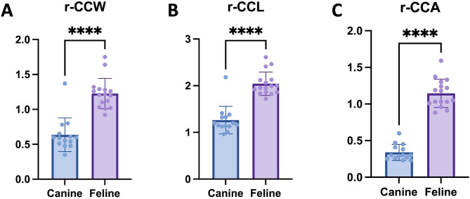
Background: This study aims to investigate the anatomical differences in the anterior segment of the eyes between dogs and cats using ultrasound biomicroscopy (UBM) to understand the higher prevalence of primary angle-closure glaucoma (PACG) in dogs compared to cats. Retrospective analysis was performed on clinical data from 16 eyes of 16 dogs and 14 eyes of 14 cats with normal eye conditions. UBM was utilized to measure nine specific parameters, including Schwalbe's Line Distance (SLD), Iridocorneal Angle (ICA), Angle-Opening Distance (AOD), and three ciliary cleft parameters: width (CCW), length (CCL), and area (CCA). To account for differences in body size, ciliary cleft parameters were adjusted accordingly.

Results: Significant anatomical differences in the anterior segment were found between the two species. Dogs had smaller values for SLD, ICA, AOD, and ciliary cleft parameters (CCW, CCL, CCA) compared to cats. Even after body-size adjustment, the rectified ciliary cleft parameters remained smaller in dogs.

Conclusion: The anatomical differences, particularly the smaller ciliary cleft and narrower drainage angles in dogs, may contribute to the higher prevalence of PACG in this species. Conversely, the larger ciliary cleft in cats may explain the lower occurrence of primary glaucoma in cats.

Keywords: Ciliary cleft; Comparative analysis; Glaucoma; Iridocorneal angle (ICA); Ultrasound biomicroscopy (UBM).


Conflict of interest statement

Declarations. Ethics approval and consent to participate: All animal owners or their representatives provided written informed consent for their pets' enrollment in the study, including the procedures and therapies undertaken, as well as for the publication of data and images derived from the study. Consent for publication: Not applicable Competing interests: The authors declare no competing interests.

Figures

Fig. 1
Fig. 1
Analysis of Iridocorneal Angle Parameters. A. Schwalbe’s Line Distance (SLD) comparison between dogs and cats shows that the mean SLD in dogs was 2.306 ± 0.1539 mm, ranging from 1.980 to 2.510 mm, whereas in cats, the mean SLD was 3.089 ± 0.2143 mm, with a range from 2.630 to 3.410 mm. B. The Angle-Opening Distance (AOD) comparison reveals that in dogs, the mean AOD was 0.9908 ± 0.3474 mm, with values ranging from 0.6200 to 1.810 mm, while in cats, the mean AOD was 1.833 ± 0.3874 mm, ranging from 1.340 to 2.580 mm. C. The Iridocorneal Angle (ICA) comparison shows that the mean ICA in dogs was 15.56 ± 3.241 degrees, ranging from 10.40 to 22.00 degrees, whereas in cats, the mean ICA was significantly larger, at 30.27 ± 3.954 degrees, with a range from 23.73 to 37.00 degrees. For ease of interpretation of our results, statistical significance is indicated as follows: not significant (ns), p < 0.05 (*), p < 0.01 (**), p < 0.001 (***), and p < 0.0001 (****)
Fig. 2
Fig. 2
Analysis of Ciliary Cleft Parameters. A. Ciliary Cleft Width (CCW) comparison between dogs and cats shows that the mean CCW in dogs was 0.6357 ± 0.2544 mm, ranging from 0.3800 to 1.450 mm, whereas in cats, the mean CCW was 1.227 ± 0.2369 mm, with a range from 0.8400 to 1.870 mm. B. The Ciliary Cleft Length (CCL) comparison reveals that in dogs, the mean CCL was 1.259 ± 0.2662 mm, with values ranging from 0.9400 to 2.050 mm, while in cats, the mean CCL was 2.039 ± 0.2518 mm, ranging from 1.660 to 2.480 mm. C. The Ciliary Cleft Area (CCA) comparison shows that the mean CCA in dogs was 0.3343 ± 0.09171 mm², ranging from 0.2200 to 0.5300 mm², whereas in cats, the mean CCA was significantly larger, at 1.149 ± 0.2388 mm², with a range from 0.7300 to 1.540 mm². For ease of interpretation of our results, statistical significance is indicated as follows: not significant (ns), p < 0.05 (*), p < 0.01 (**), p < 0.001 (***), and p < 0.0001 (****)
Fig. 3
Fig. 3
Analysis of Rectified Ciliary Cleft Parameters. A. Rectified Ciliary Cleft Width (r-CCW) comparison between dogs and cats shows that the mean r-CCW in dogs was 0.6364 ± 0.2410 mm, ranging from 0.3500 to 1.370 mm, whereas in cats, the mean r-CCW was 1.227 ± 0.2181 mm, with a range from 0.9200 to 1.750 mm. B. The Rectified Ciliary Cleft Length (r-CCL) comparison reveals that in dogs, the mean r-CCL was 1.264 ± 0.2937 mm, with values ranging from 0.9600 to 2.180 mm, while in cats, the mean r-CCL was 2.043 ± 0.2513 mm, ranging from 1.720 to 2.610 mm. C. The Rectified Ciliary Cleft Area (r-CCA) comparison shows that the mean r-CCA in dogs was 0.3386 ± 0.1088 mm², ranging from 0.2300 to 0.6000 mm², whereas in cats, the mean r-CCA was significantly larger, at 1.145 ± 0.1917 mm², with a range from 0.8800 to 1.590 mm². For ease of interpretation of our results, statistical significance is indicated as follows: not significant (ns), p < 0.05 (*), p < 0.01 (**), p < 0.001 (***), and p < 0.0001 (****)
Fig. 4
Fig. 4
Comparison of Ciliary Clefts in Dogs and Cats. A. Iridocorneal angle in dogs. B. Iridocorneal angle in cats. The comparison of the two images shows that cats have a wider ciliary cleft than dogs
Fig. 5
Fig. 5
UBM Examination Parameters. A. Iridocorneal angle parameters: Schwalbe’s Line Distance (SLD) was measured from the borderline of the cornea and sclera to the anterior lens capsule. The Iridocorneal Angle (ICA) was identified at the junction of the sclera, cornea, and iris base, with measurements taken between the inner scleral layer and the outer iris root. The Angle-Opening Distance (AOD) was measured perpendicularly from Descemet’s membrane to the anterior iris surface. B. Ciliary cleft parameters: Ciliary Cleft Width (CCW) was measured from the pectinate ligament’s outer layer to the iris root’s upper surface. Ciliary Cleft Length (CCL) was measured from the angle recess to the midpoint of the CCW. C. Ciliary Cleft Area (CCA) was calculated by tracing the inner scleral surface from the angle recess to its boundary and the superior iris root up to the CCW, providing insights into the CC’s anatomical structure

Similar articles

References

    1. Miller PE, Bentley E. Clinical signs and diagnosis of the canine primary glaucomas. Vet Clin North Am Small Anim Pract. 2015;45(6):1183–212. vi. - PMC - PubMed
    1. PE M. The Glaucomas2008. 230–57. p.
    1. Maggs DMP, Ofri R, editors. Slatter’s fundamentals of veterinary ophthalmology. St. Louis: Elsevier; 2018.
    1. Gelatt KN, MacKay EO. Prevalence of the breed-related glaucomas in pure-bred dogs in North America. Vet Ophthalmol. 2004;7(2):97–111. - PubMed
    1. Gelatt KN, MacKay EO. Secondary glaucomas in the dog in North America. Vet Ophthalmol. 2004;7(4):245–59. - PubMed

Publication types

MeSH terms

LinkOut - more resources