. 2025 Mar 18;26(1):275.
doi: 10.1186/s12891-025-08512-3.

Optimizing uni-compartmental knee arthroplasty: the impact of preoperative planning and arithmetic hip-knee-ankle angle

Affiliations

Optimizing uni-compartmental knee arthroplasty: the impact of preoperative planning and arithmetic hip-knee-ankle angle

Changquan Liu et al. BMC Musculoskelet Disord. .

Abstract

Purpose: The purpose of this study was to evaluate whether the combination of preoperative planning software combined with arithmetic hip-knee-ankle angle (aHKA) can help patients who underwent uni-compartmental knee arthroplasty (UKA) recover the constitutional alignment of the lower limb, obtain a better prosthetic position, and achieve better early patient-reported outcome measurements (PROMs).

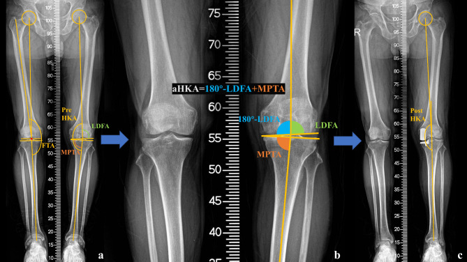
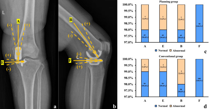
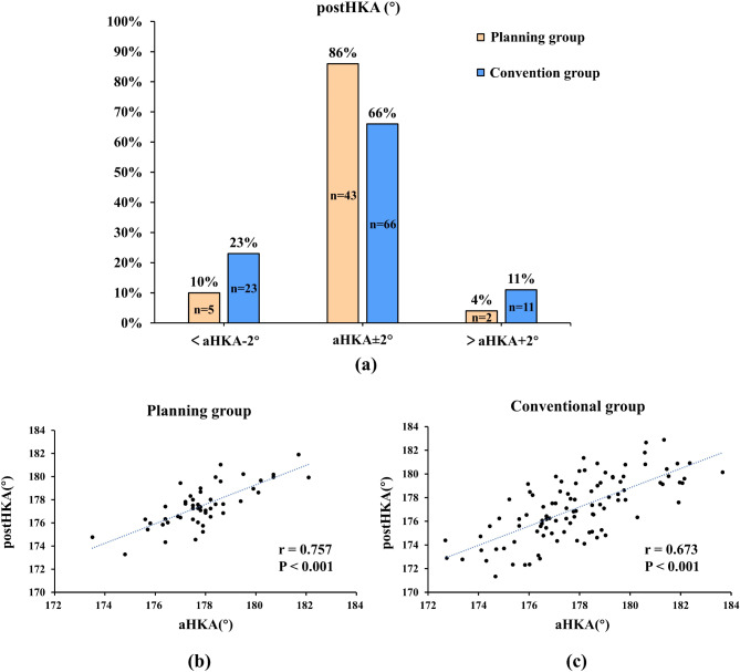
Methods: A total of 150 patients who underwent UKA (planning group: 50 patients using the preoperative planning software; conventional group: 100 patients using the conventional method) were included in the study. The aHKA was defined as 180° + mechanical medial proximal tibial angle (MPTA) - mechanical distal lateral femoral angle (LDFA). All patients in the planning group underwent UKA according to the planning software with the planned lower limb alignment of aHKA. All patients were divided into three groups: constitutional alignment group (postoperative HKA (post-HKA): aHKA ± 2.0°); overcorrection group (post-HKA > aHKA + 2.0°); under-correction group (post-HKA < aHKA - 2.0°). Comparisons between the planning and conventional groups were conducted: (1) the proportion of post-HKA restored to constitutional alignment group; (2) the postoperative prosthesis position parameter based on the guideline of the Oxford group; (3) the American Knee Society scores (KSS) at six months after surgery.

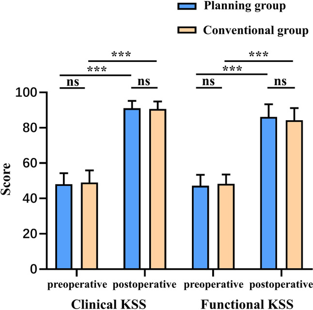
Results: The proportion of the constitutional alignment group in the planning group was higher than that in the conventional group (86% vs. 66%) (p = 0.033). There was no significant difference in postoperative prosthesis position parameters between the two groups. No significant difference was found between the KSS clinical score (91.02 ± 4.20 vs. 90.61 ± 4.24) and KSS functional score (86.10 ± 7.23 vs. 84.30 ± 6.82) in six months after surgery between the planning and conventional groups.

Conclusion: Patients who underwent UKA using preoperative planning software in combination with aHKA were able to recover a higher proportion of the constitutional alignment than those with the conventional method. In addition, the planning group could achieve similar postoperative prosthesis position and short-term PROMs compared to the conventional group.

Clinical trial number: Not applicable.

Keywords: Arithmetic hip-knee-ankle angle (aHKA); Constitutional alignment.; Lower limb alignment; Preoperative planning software; Uni-compartmental knee arthroplasty (UKA).


Conflict of interest statement

Declarations. Ethics approval and consent to participate: All procedures performed in studies involving human participants were in accordance with the ethical standards of the institutional and national research committee and with the 1964 Helsinki Declaration and its later amendments or comparable ethical standards. The study was approved by the institutional review board of the China-Japan Friendship Hospital (approval number 2020–50-k28). The written informed consent was obtained from all individual participants included in the study. Consent for publication: Not applicable. Competing interests: The authors declare no competing interests.

Figures

Fig. 1
Fig. 1
Pattern diagram of radiographic parameter measurements (a-c). (a) The preoperative weight-bearing full-length radiograph. The preoperative hip–knee–ankle angle (pre-HKA), femoral tibial angle (FTA), mechanical lateral distal femoral angle (LDFA), and mechanical proximal tibial angle (MPTA) were measured; (b) Partial magnification of the preoperative weight-bearing full-length radiograph. The arithmetic hip-knee-ankle angle (aHKA) was calculated as 180° + MPTA − LDFA; (c) The postoperative weight-bearing full-length radiograph. The postoperative HKA (post-HKA) was measured
Fig. 2
Fig. 2
Pattern diagram of postoperative prosthetic parameter measurements (a-b). (a) The postoperative anterior-posterior (AP) knee radiograph. The coronal angle of the femoral prosthesis varus/valgus (femoral angle A) and the coronal angle of the tibial prosthesis varus/valgus (tibial angle E) were measured; (b) The postoperative lateral knee radiograph. The sagittal angle of the femoral prosthesis flexion/extension (femoral angle B) and posterior tibial prosthesis slope (tibial angle F) were measured; (c) Distribution of postoperative prosthesis position in the planning group. 47 (94%) patients achieved acceptable prosthesis position while 3 (6%) patients were in poor prosthesis position, including 1 (2%) patient with an angle A greater than 10°, 1 (2%) patient with an angle E greater than 5°, and 1 (2%) patient with an angle B greater than 15°; (d) Distribution of postoperative prosthesis position in the conventional group. 96 (96%) patients achieved acceptable prosthesis position while 4 (4%) patients were in poor prosthesis position, including 1(1%) patient with an angle A greater than 10°, 1 (1%) patient with an angle E greater than 5°, and 2 (2%) patient with an angle B greater than 15°
Fig. 3
Fig. 3
Usage steps of the full-length radiograph software (a-d). (a) Step 1, import the full-length radiograph; (b) Step 2, manually mark the key points, automatically connect the axes and calculate the angles; (c) Step 3, manually preset the angle of hip–knee–ankle angle (HKA), and automatically display the original HKA, femoral tibial angle (FTA) and medial joint space width (mJSW); (d) Step 4, recognize and segment the tibia and fibula, automatically rotate the tibia and fibula to the preset HKA, and display HKA, FTA, and mJSW after rotation
Fig. 4
Fig. 4
Usage steps of the full-length computed tomography (CT) software (a-d). (a) Step 1, import the full-length CT and segment the full-length CT of the target side by artificial intelligence method; (b) Step 2, manually mark the key points, automatically connect the axes and calculate the angles; (c) Step 3, manually preset the angle of hip–knee–ankle angle (HKA), and automatically display the original HKA, femoral tibial angle (FTA) and medial joint space width (mJSW); (d) Step 4, automatically rotate the tibia and fibula to the preset HKA and display HKA, FTA, and mJSW after rotation
Fig. 5
Fig. 5
Measurement of intraoperative femoral grinding depth (d2) and tibial osteotomy thickness (d3) (a-f). (a) Tibial longitudinal osteotomy process; (b) Tibial horizontal osteotomy process; (c) Femoral grinding process. The depth of femur grinding is recorded as d2; (d) Tibial osteotomy block. The marked point is the lowest point of tibial wear; (e) Measuring instrument. The range is 0–10 mm, with an accuracy of 0.1 mm; (f) Using the measuring instrument to measure the thickness of the tibial osteotomy block (the thickness at the lowest point of tibial wear). The thickness of tibial osteotomy (d3) = tibial osteotomy block thickness + blade thickness (1.2 mm)
Fig. 6
Fig. 6
Surgical pattern diagram of the planning group (a-c). (a) Preoperative pattern diagram. The preoperative planned medial joint space width is d1 (corresponding to the planned alignment of arithmetic hip-knee-ankle angle (aHKA)); (b) Intraoperative pattern diagram. The depth of femoral grinding during surgery is d2, and the tibial osteotomy thickness is d3; (c) Postoperative pattern diagram. The thickness of the tibial prosthesis is d4, the thickness of the distal femoral prosthesis is d5, and the thickness of the polyethylene bearing is d6. During the operation, the d6 is adjusted to meet the requirement of d1 + d2 + d3 = (d4 + d5 + d6) ± 1 mm; (d) The planned medial joint space width (d1) and tibial osteotomy thickness (d3). The mean ± standard deviation (SD) of d1 and d3 were 4.82 ± 0.75 mm and 3.37 ± 0.64 mm; (e) Femoral grinding depth (d2). The number of patients with d2 of 2/3/4/5 mm is 13 (26.0%)/28 (56.0%)/7 (14.0%)/2 (4.0%), respectively; (f) The total space width (d1 + d2 + d3 or d4 + d5 + d6) and space width difference (Δd= (d1 + d2 + d3) - (d4 + d5 + d5)). d1 + d2 + d3 = 11.15 ± 0.87 mm, d4 + d5 + d6 = 10.46 ± 0.80 mm, and Δd is 0.69 ± 0.31 mm
Fig. 7
Fig. 7
The relationship between postoperative hip–knee–ankle angle (post-HKA) and arithmetic hip-knee-ankle angle (aHKA) in the planning group and the conventional group. (a) The postoperative alignment grouping. Constitutional alignment group (post-HKA: aHKA ± 2.0°), overcorrection alignment group (post-HKA > aHKA + 2.0°), under-correction alignment group (post-HKA < aHKA − 2.0°); (b) Correlation analysis in the planning group; (c) Correlation analysis in the conventional group
Fig. 8
Fig. 8
The American Knee Society scores (KSS) before and six months after surgery in the preoperative planning group and the conventional group. There was no significant difference in the preoperative KSS clinical score (48.08 ± 6.25 vs. 49.04 ± 6.87), postoperative KSS clinical score (91.02 ± 4.20 vs. 90.61 ± 4.24), preoperative KSS functional score (47.20 ± 6.16 vs. 48.30 ± 5.23), and postoperative KSS functional score (86.10 ± 7.23 vs. 84.30 ± 6.82) between the two groups. Both groups showed significant improvement in KSS scores six months post-surgery

Similar articles

References

    1. Slaven SE, Cody JP, Sershon RA, Ho H, Hopper RH Jr., Fricka KB. Alignment in medial Fixed-Bearing unicompartmental knee arthroplasty: the limb has a leg up on the component. J Arthroplasty. 2021;36(12):3883–7. 10.1016/j.arth.2021.08.015. - PubMed
    1. Liu C, Chen H, Ge J, Huang C, Zhang Q, Guo W. Predicting valgus malalignment after mobile-bearing UKA using a new method: the arithmetic HKA of the arthritic knee. Arch Orthop Trauma Surg. 2023;143(10):6381–91. 10.1007/s00402-023-04921-y. - PubMed
    1. Itou J, Kuwashima U, Itoh M, Okazaki K. Perioperative varus alignment does not affect short-term patient-reported outcome measures following mobile-bearing unicompartmental knee arthroplasty. J Orthop Surg Res. 2022;17(1):98. 10.1186/s13018-022-02999-5. - PMC - PubMed
    1. Crawford DA, Berend KR, Thienpont E. Unicompartmental knee arthroplasty: US and global perspectives. Orthop Clin North Am. 2020;51(2):147–59. 10.1016/j.ocl.2019.11.010. - PubMed
    1. Grant AL, Doma KD, Hazratwala K. Determination of the accuracy of navigated kinematic unicompartmental knee arthroplasty: A 2-Year Follow-Up. J Arthroplasty. 2017;32(5):1443–52. 10.1016/j.arth.2016.11.036. - PubMed

LinkOut - more resources