. 2025 Mar 18;23(1):221.
doi: 10.1186/s12951-025-03201-8.

The immunogenic potential of an optimized mRNA lipid nanoparticle formulation carrying sequences from virus and protozoan antigens

Affiliations

The immunogenic potential of an optimized mRNA lipid nanoparticle formulation carrying sequences from virus and protozoan antigens

Renata S Fernandes et al. J Nanobiotechnology. .

Abstract

Background: Lipid nanoparticles (LNP) are a safe and effective messenger RNA (mRNA) delivery system for vaccine applications, as shown by the COVID-19 mRNA vaccines. One of the main challenges faced during the development of these vaccines is the production of new and versatile LNP formulations capable of efficient encapsulation and delivery to cells in vivo. This study aimed to develop a new mRNA vaccine formulation that could potentially be used against existing diseases as well as those caused by pathogens that emerge every year.

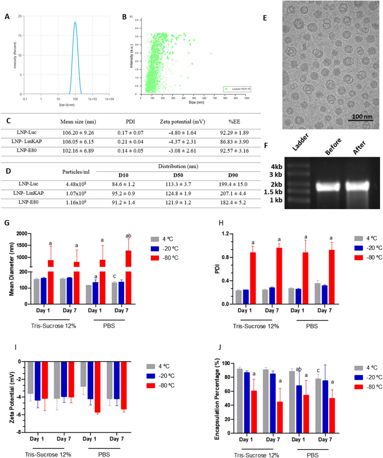
Results: Using firefly luciferase (Luc) as a reporter mRNA, we evaluated the physical-chemical properties, stability, and biodistribution of an LNP-mRNA formulation produced using a novel lipid composition and a microfluidic organic-aqueous precipitation method. Using mRNAs encoding a dengue virus or a Leishmania infantum antigen, we evaluated the immunogenicity of LNP-mRNA formulations and compared them with the immunization with the corresponding recombinant protein or plasmid-encoded antigens. For all tested LNP-mRNAs, mRNA encapsulation efficiency was higher than 85%, their diameter was around 100 nm, and their polydispersity index was less than 0.3. Following an intramuscular injection of 10 µg of the LNP-Luc formulation in mice, we detected luciferase activity in the injection site, as well as in the liver and spleen, as early as 6 h post-administration. LNPs containing mRNA encoding virus and parasite antigens were highly immunogenic, as shown by levels of antigen-specific IgG antibody as well as IFN-γ production by splenocytes of immunized animals that were similar to the levels that resulted from immunization with the corresponding recombinant protein or plasmid DNA.

Conclusions: Altogether, these results indicate that these novel LNP-mRNA formulations are highly immunogenic and may be used as novel vaccine candidates for different infectious diseases.

Keywords: Biodistribution; Dengue; IFNγ; IgG; Leishmaniasis; Lipid nanoparticles; mRNA vaccine.


Conflict of interest statement

Declarations. Competing interests: The authors declare no competing interests.

Figures

Fig. 1
Fig. 1
Physical–chemical characterization of LNP-mRNA. Size distribution of LNP-mRNA was measured by a dynamic light scattering (DLS) and b Nanoparticle Tracking Analysis (NTA). c Mean diameter, polydispersity index (PDI), zeta potential, encapsulation efficiency (%EE) for different mRNA loaded LNP d NTA results: mean size, distribution (D10, D50, D90), and particle concentration of LNP-Luc, LNP-LinKAP, and LNP-E80 e Cryo-TEM images of LNP-mRNA by plunge freezing technique f Analysis of Luc-mRNA in agarose gel in denaturing conditions: 1- mRNA-Luc before encapsulation and 2- mRNA-Luc after encapsulation into LNP. g Mean diameter, h PDI, i zeta potential, and j encapsulation percentage of LNP-mRNA prepared with PBS or Tris–HCl + Sucrose 12% and stored at 4 °C, -20 °C, or − 80 °C. Data are expressed by the mean (n = 3) ± standard deviation of the mean. All data were analyzed using a one-way ANOVA analysis of variance followed by Tukey's post-test. Different letters indicate significant differences among groups (p < 0.05)
Fig. 2
Fig. 2
Delivery of Luc-mRNA in vitro and in vivo. A HEK293T cells were transfected with naked Luc-mRNA or 3, 6, or 9 ug with Luc-mRNA encapsulated in LNPs. Twenty-four hours after transfection, cells were collected, and total protein extracts were prepared and used in luciferase assays. B Cytotoxicity evaluation of LNP-Luc using sulforhodamine B (SRB) assay. HEK293T cells were treated with different amounts of free or encapsulated mRNA for 24 h. DMSO was used as a positive control for cellular toxicity. C In vivo BLI of LNP-Luc in mice. C Female BALB/c mice were inoculated with 10 ug of LNP-Luc via IM and subjected to IVIS Spectrum imaging at the indicated times after administration. D Tissue distribution of LNP-Luc in mice, and E quantification of tissue distribution of LNP-Luc in mice 24 h after administration. F Quantification of luminescence expressed in photons per second (p/s) following IM inoculation
Fig. 3
Fig. 3
In vitro expression of LNP mRNAs and in vivo immunogenicity A mRNA from two different antigens (E80 dengue virus serotype 3 and LinKAP from Leishmania) were in vitro synthesized and HEK293T cells were transfected with both LNP-mRNAs respectively. Forty-eight hours after transfection, cells were collected, and a Western blot was performed with B culture supernatant from cells transfected with E80 or C total protein extract from cells transfected with LinKAP. D Female C57BL/6 mice were immunized IM with 10 µg of LNP-E80 or LNP-LinKAP, naked mRNA or empty-LNP, with a prime-boost or prime-boost-boost regimen at a 3-week interval. Serum was collected on days 20, 41 and 62 to analyze specific total IgG. E Total IgG titers after mice immunization with LNP-E80 after the boost (p < 0.0001). Thirty days after the last immunization, mice spleens were collected and used in splenocyte cultures with the appropriate stimuli. F IFN-γ production in mice immunized with LNP-E80 (****p < 0.0001, ***p < 0.001). G Total IgG titers after mice immunization with LNP-LinKAP (p < 0.0001). H IFN-γ production in mice immunized with LNP-LinKAP (**p = 0.0001, *p = 0.0001). Statistical analysis was performed with 2-way ANOVA with Turkey’s multiple comparisons test
Fig. 4
Fig. 4
Evaluation of innate immune response. a Splenocytes were isolated from Balb/c naïve mice and stimulated with either empty or mRNA-loaded, or naked mRNA. Sixteen hours post-incubation, cytokine levels were measured using the CBA Mouse Th1/Th2/Th17 kit. b Cytokine production in response to LNP with or without mRNA and SM-102. Mouse splenocyte cultures were stimulated with LNP, and cytokine levels were measured 16 h after incubation for TNF, IFN-γ, IL-6, IL-10, IL-2, IL-4, and IL-17

References

    1. Fire A, Xu S, Montgomery MK, Kostas SA, Driver SE, Mello CC. Potent and specific genetic interference by double-stranded RNA in Caenorhabditis elegans. Nature. 1998;391(6669):806–11. - PubMed
    1. Kon E, Elia U, Peer D. Principles for designing an optimal mRNA lipid nanoparticle vaccine. Curr Opin Biotechnol. 2022;73:329–36. - PMC - PubMed
    1. Hassett KJ, Benenato KE, Jacquinet E, Lee A, Woods A, Yuzhakov O, Himansu S, DeterLing J, Geilich BM, Ketova T, Mihai C, Lynn A, McFadyen I, Moore MJ, Senn JJ, Stanton MG, Almarsson O, Ciaramella G, Brito LA. Optimization of lipid nanoparticles for intramuscular administration of mrna vaccines. Mol Ther Nucleic Acids. 2019;15:1–11. - PMC - PubMed
    1. Jackson NAC, Kester KE, Casimiro D, Gurunathan S, DeRosa F. The promise of mRNA vaccines: a biotech and industrial perspective. NPJ Vaccines. 2020;5:11. - PMC - PubMed
    1. Pilkington EH, Suys EJA, Trevaskis NL, Wheatley AK, Zukancic D, Algarni A, Al-Wassiti H, Davis TP, Pouton CW, Kent SJ, Truong NP. From influenza to COVID-19: lipid nanoparticle mRNA vaccines at the frontiers of infectious diseases. Acta Biomater. 2021;131:16–40. - PMC - PubMed

MeSH terms

LinkOut - more resources