. 2025 Mar 18;20(1):292.
doi: 10.1186/s13018-025-05557-x.

Fibroblast IRF7-mediated chondrocyte apoptosis affects the progression of collapse in steroid-induced osteonecrosis of the femoral head

Affiliations

Fibroblast IRF7-mediated chondrocyte apoptosis affects the progression of collapse in steroid-induced osteonecrosis of the femoral head

Han Longfei et al. J Orthop Surg Res. .

Abstract

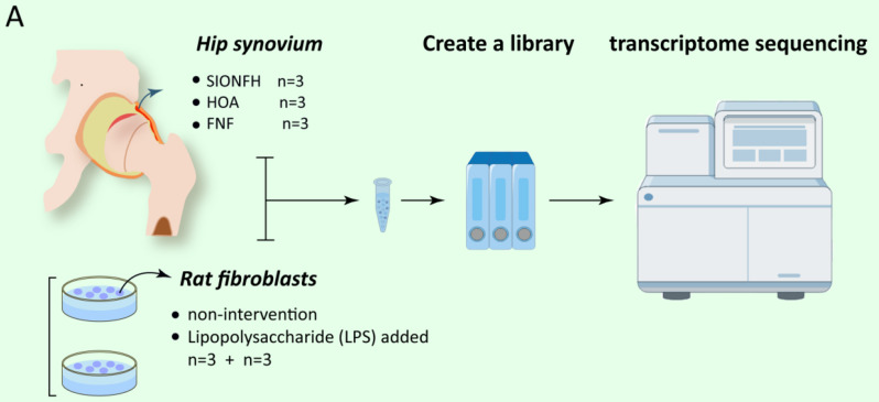
Purpose: The objective of this study was to identify potential genes implicated in the "peri-collapse" synovium of osteonecrosis of the femoral head through coding gene sequencing and to further clarify their specific mechanisms via in vitro experiments.

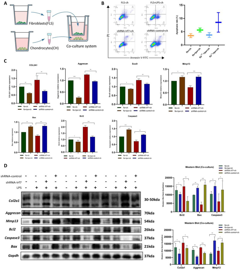
Methods: Steroid-induced osteonecrosis of the femoral head (SIONFH) (n = 3), femoral neck fracture (FNF) (n = 3), and hip osteoarthritis (HOA) (n = 3) Synovial tissue of the hip joint was collected in total hip arthroplasty. A cellular model of SIONFH constructed from rat synovial fibroblasts by lipopolysaccharide intervention. Lentiviral technology was used to construct a model for fibroblast knockout of the Irf7 gene. HE was used to compare the characteristics of synovial tissue damage, and immunofluorescence and immunohistochemistry were used to compare the expression levels of VIM, IRF7, and IFNα. PCR, WB, and IF were used to examine Irf7 knockdown efficiency, chondrocyte proliferation (Col2a1, Aggrecan, Sox9), cartilage matrix degradation (Mmp13), and apoptosis (Bcl2, Bax, and Caspase3) expression under co-culture conditions. Crystalline violet staining was used to observe the migration rate of fibroblasts, and flow cytometry was used to detect the apoptosis level of chondrocytes under co-culture conditions.

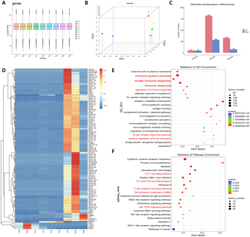
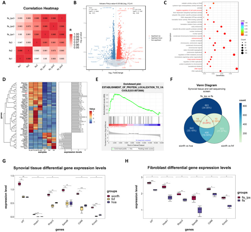
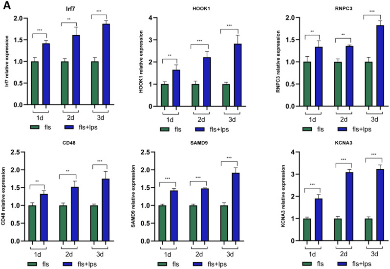
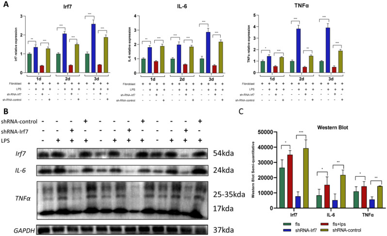
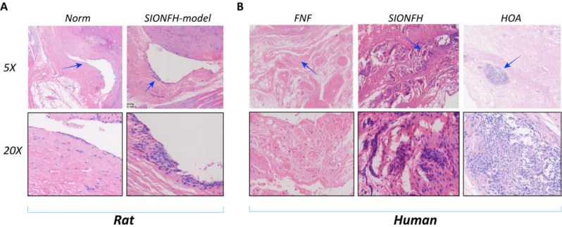
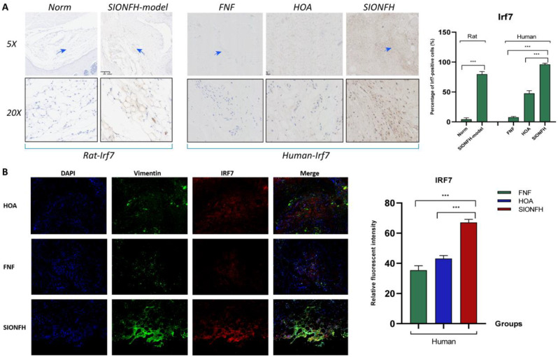
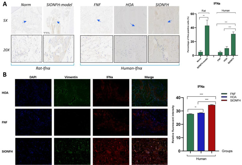
Results: Transcriptome sequencing of synovial tissue and fibroblasts ultimately screened for six differential genes, HOOK1, RNPC3, KCNA3, CD48, IRF7, SAMD9. Compared to FNF and HOA, synovial inflammatory cell recruitment and synovial hyperplasia were more pronounced in SIONFH. IF and IHC confirmed high expression of IRF7 and IFNα in the synovium of SIONFH. PCR and WB results suggested that fibroblasts highly expressed Irf7, Hook1, Rnpc3, Kcna3, Cd48, Samd9, Il-6, and Tnfα after lipopolysaccharide intervention, and the expression levels of Il-6 and Tnfα were significantly reduced after knockdown of Irf7 (P < 0.001). In the co-culture system, fibroblasts intervened with lipopolysaccharide significantly promoted chondrocyte apoptosis, the rate of cartilage matrix degradation, while inhibiting the level of chondrocyte proliferation, and this result was significantly reversed in Irf7 knockout fibroblasts. This was supported by flow cytometry results.

Conclusions: IRF7, HOOK1, RNPC3, KCNA3, CD48, and SAMD9 as potential genes affecting the progression of SIONFH collapse. Irf7 mediates the fibroblast inflammatory response and affects the collapse process of SIONFH by influencing chondrocyte apoptosis. Thus, intervention in IRF7 holds promise as one of the key targets for reversing the collapse process of SIONFH.

Keywords: Apoptosis; Collapse; Gene expression profiles; Steroid-induced osteonecrosis of the femoral head; Synovial fibroblasts.


Conflict of interest statement

Declarations. Ethics approval and consent to participate: This study was approved by the board of research ethics in The Third Affiliated Hospital of Guangzhou University of Chinese Medicine (Approval No. PJ-XS-20230523-002). All patients give informed consent. Consent for publication: All authors of this manuscript agree with the views expressed in this article and consent to publication. Competing interests: The authors declare no competing interests.

Figures

Fig. 1
Fig. 1
Schematic of RNA sequencing. In order for the results to be statistically significant, SIONFH hip synovium (n = 3),FNF hip synovium (n = 3), HOA hip synovium (n = 3), lipopolysaccharide-treated rat fibroblasts for 48 h (n = 3) and untreated cells (n = 3) were selected for RNA sequencing. This was followed by quality control, library construction and sequencing
Fig. 2
Fig. 2
Hip synovial tissue RNA sequencing quality control and results. [A] Gene expression boxplot reaction consistency of 9 samples. The results showed that all samples were in good agreement. [B] The PCA clustering results of 9 samples showed that the intra-group differences of samples in FNF, HOA and SIONFH groups were small, and the inter-group differences between SIONFH and FNF and HOA groups were large. [C] The number of up- and down-regulated genes in HOA vs. FNF, SIONFH vs. FNF, and SIONFH vs. HOA groups compared. Red color represents the number of up-regulated genes and blue color represents the number of down-regulated genes. [D] Heatmap of differential genes from RNA sequencing of synovial tissues of SIONFH, HOA, and FNF groups showed that IRF7, IFNα, and IG (immunoglobulin) family genes were significantly highly expressed in the synovial tissues of SIONFH (P < 0.05). HMCN2 was significantly highly expressed in the synovial tissues of HOA (P < 0.05). genes such as CHI3L2, DMKN, and other genes were significantly highly expressed in the synovial tissues of SIONFH. FNF synovial tissues (P < 0.05). [E] GO enrichment analysis of SIONFH vs. FNF vs. HOA differential genes showed that the differential genes in SIONFH synovium were mainly enriched in the process of immune system, innate immune response, regulation of immune response, B-cell receptor signaling pathway, and positive regulation of B-cell activation, etc. The results showed that SIONFH synovium had a high level of expression of CHI3L2, DMKN and other genes (P < 0.05). [F] KEGG enrichment of SIONFH vs. FNF vs. HOA differential genes showed that SIONFH differential genes were enriched in Th17 cell differentiation, Th1 and Th2 cell differentiation, T-cell receptor signaling pathway, cell adhesion molecules (CAMs), and Jak-STAT signaling pathway
Fig. 3
Fig. 3
Figure 3 HOOK1, RNPC3, KCNA3, CD48, IRF7, and SAMD9 are highly expressed in SIONFH synovium during the “peri-collapse phase”. [A] Heatmap of correlation coefficients of RNA sequencing samples from 5ug/ml lipopolysaccharide-treated fibroblasts (the higher the correlation coefficient between the samples, the closer their expression patterns). The results showed that the consistency of the samples in the lipopolysaccharide intervention group was better, and the fibroblast control group sample 3 was more different from samples 1 and 2, but the subsequent analysis was still performed considering its value of 0.947, which is close to 1. [B] Volcano plot of differential genes from RNA sequencing of fibroblasts, red represents up-regulated differential genes in lipopolysaccharide-treated fibroblasts and blue represents down-regulated differential genes. (P-value < 0.05 and |log2 FC| > 1.5). [C] Bubble plots of GO enrichment results for differential genes in lipopolysaccharide-treated fibroblasts showing enrichment in negative regulation of cell cycle process, endosomal part, Golgi vesicle transport, DNA repair. [D] Heatmap of RNA sequencing differential genes in lipopolysaccharide-treated fibroblasts showing Irf7, Tph1, Calcrl, Spsb2 are highly expressed in the lipopolysaccharide -treated group, and genes such as Rcan2, Rps9, and Apln are lowly expressed in the lipopolysaccharide-treated group. Dendrograms of correlation are shown on the left, and histograms of fold difference are shown on the right. [E] GSEA plot of RNA sequencing enrichment analysis of lipopolysaccharide-treated fibroblasts, which showed significant enrichment in the ESTABLISHMENT OF PROTEIN LACALIZATION TO VACUOLE entry. This suggests that protein transport and translocation may play a key role. [F] Venn diagram of RNA sequencing of SIONFH hip synovial tissue with RNA sequencing of differential genes in lipopolysaccharide -treated fibroblasts. Six differential genes were finally screened, namely HOOK1, RNPC3, KCNA3, CD48, IRF7, and SAMD9. [G] Differential expression of HOOK1, RNPC3, KCNA3, CD48, IRF7, and SAMD9 in synovial tissues of hip joints (Note: * indicates P < 0.05; ** indicates P < 0.01; *** indicates P < 0.001). Different colors represent different samples. [H] Differential expression of Hook1, Rnpc3, Kcna3, Cd48, Irf7, and Samd9 in RNA sequencing of lipopolysaccharide -treated fibroblasts (Note: * indicates P < 0.05; ** indicates P < 0.01; *** indicates P < 0.001). Different colors represent different samples
Fig. 4
Fig. 4
In vitro experiments showed that LPS significantly up-regulated the expression of Irf7, Hook1, Rnpc3, Cd48, Samd9, Kcna3 in fibroblasts. The experiments included a fibroblast control group and a 5ug/ml LPS intervention group, and the cells were collected on days 1,2,3 for PCR validation, respectively. (Note: * indicates P < 0.05; ** indicates P < 0.01; *** indicates P < 0.001). Significance analysis was performed using Independent Samples t-Test
Fig. 5
Fig. 5
Il-6, Tnfα significantly downregulated in fibroblasts knocked down for Irf7. [A] To verify the efficiency of lentiviral transfection to knockdown Irf7, we performed RT-qPCR experiments. The experiment was divided into four groups, fibroblast control group, 5ug/ml lipopolysaccharide treatment group, shRNA-Irf7 transfection group, and shRNA-control transfection group. Cells in each group were cultured at 37 °C under 5% CO2 for 1,2,3 days, after which PCR experiments were performed for verification. In addition, in order to explore the downstream genes of Irf7, we also detected Il-6, Tnfα by PCR experiments. (Note: One-way ANOVA was used for multiple comparisons; * indicates P < 0.05; ** indicates P < 0.01; *** indicates P < 0.001). [B] To verify the expression of relevant proteins after knockdown of Irf7, we performed Western Blot experiments. The experimental grouping was as above. We examined the expression of Irf7, Il-6, Tnfα proteins, the experiment was set up with 3 groups of replicates, and the protein molecular weights were labeled on the right side of the bands. [C] In order to quantitatively represent the expression of Irf7, Il-6, Tnfα proteins, we semi-quantitatively analyzed the bands and performed statistical plots. (Note: * indicates P < 0.05; ** indicates P < 0.01)
Fig. 6
Fig. 6
Knockdown of Irf7 inhibits the level of cell migration in fibroblasts. [A] To verify the expression level of Irf7 gene after lentiviral transfection, we performed immunofluorescence (IF) experiments. The experiment was divided into four groups, namely, fibroblast control group, 5ug/ml lipopolysaccharide treatment group, shRNA-Irf7 group, and shRNA-control group. Cells were cultured at 37 °C under 5% CO2 for 48 h for IF experiments. In order to quantitatively analyze the efficiency of knockdown of Irf7, we semi-quantitatively analyzed the IF results. (Note: One-way ANOVA was used for multiple samples; ** indicates P < 0.01; *** indicates P < 0.001). [B] To detect the migratory ability of fibroblasts, we performed experiments and stained with crystal violet. The experimental grouping was as above. The experimental method was to seed equal numbers of fibroblasts into the upper chamber of Transwell, and only serum-containing medium was added to the lower chamber. 48 h later, the fibroblasts in the lower chamber were subjected to crystal violet staining and the number of cells was counted. (Note: One-way ANOVA was used for multiple samples; *** indicates P < 0.001)
Fig. 7
Fig. 7
Synovitis manifestations are more prominent in the SIONFH hip synovium. [A] To compare the level of synovitis in SIONFH disease, we built a SIONFH rat model and used HE staining to compare the level of inflammatory response in the synovium. Arrowheads stand in for magnified areas. [B] To compare the level of synovial inflammation in different diseases, we took hip synovium from femoral neck fracture (FNF), hip osteoarthritis (HOA), and hormonal osteonecrosis of the femoral head (SIONFH) for HE staining. Arrowheads stand in for magnified areas
Fig. 8
Fig. 8
IRF7 is significantly overexpressed in the synovium of SIONFH hip joints. [A] In order to clarify the expression of IRF7 in the synovium of SIONFH hips, we performed immunohistochemistry (IHC) experiments for IRF7 in the synovium of hips from SIONFH-Rat and in the synovium of hips from patients with FNF, HOA, and SIONFH. Arrowheads stand in for magnified areas, and we also counted and plotted the number of positive cells for IRF7. [B] Based on the above purpose, we also performed immunofluorescence (IF) experiments of IRF7 and VIM on the synovium of the hip joints of patients with FNF, HOA, and SIONFH, and we also statistically analyzed and graphed the average fluorescence intensity of the synovial samples of the three groups. (Note: One-way ANOVA was used for multiple group comparisons; *** indicates P < 0.001)
Fig. 9
Fig. 9
IFNα downstream of IRF7 is significantly overexpressed in SIONFH hip synovium. [A] IFNα is a downstream protein of IRF7, therefore, we tested IFNα. As above, we performed IHC experiments for IFNα and counted and plotted the number of positive cells. (Note: One-way ANOVA was used for multiple comparisons; * indicates P < 0.05; *** indicates P < 0.001). [B] Similarly, we performed fluorescence double staining experiments of IFNα and VIM in the synovium of patients’ hip joints, and the average fluorescence intensity was counted and plotted. (Note: One-way ANOVA was used for multiple group comparisons; *** indicates P < 0.001)
Fig. 10
Fig. 10
Knockdown of Irf7 reverses synoviocyte-mediated chondrocyte apoptosis. [A] To clarify the effect of synovial fibroblasts on chondrocytes, we established a cell co-culture device. It was divided into 4 groups, which were fibroblast control group, 5ug/ml lipopolysaccharide treatment group, shRNA-Irf7 transfection group, and shRNA-control transfection group. The cells were pretreated for 48 h, after which they were transferred into the upper chamber of the co-culture system, and the chondrocytes were transferred into the lower chamber of the co-culture system, and cultured at 37℃ with 5% CO2 for 48 h, after which they were subjected to RT-qPCR, Western Blot, and flow cytometry. [B] To verify the effect of synovial fibroblasts on chondrocytes, we used flow cytometry for detecting the level of chondrocyte apoptosis. And the results were statistically analyzed and counted. [C] To verify the effect of synovial fibroblasts on mRNA expression of chondrocytes, we used RT-qPCR for detecting the mRNA expression level of chondrocyte Col2a1, Aggrecan, Sox9, Mmp13, Bax, Bcl2, and Caspase3 indicators. Grouping as above. (Note: One-way ANOVA was used for multiple group comparisons; * indicates P < 0.05; ** indicates P < 0.01; *** indicates P < 0.001). [D] To verify the effect of synovial fibroblasts on chondrocyte apoptosis, we examined the protein expression of chondrocyte Col2a1, Aggrecan, Mmp13, Bax, Bcl2, Caspase3. The grouping was as above. In order to quantify the amount of expressed proteins, we used Image J software to analyze the bands semi-quantitatively, and performed statistical analysis and graphing. (The experiment was set up with 3 groups of replicates, and one-way ANOVA was used for multiple comparisons; * indicates P < 0.05; ** indicates P < 0.01; *** indicates P < 0.001)

Similar articles

References

    1. Zhai S, Wu R, Zhao J, Huang W, Hu W, Huang W. Effectiveness of various interventions for non-traumatic osteonecrosis: a pairwise and network meta-analysis. Front Endocrinol (Lausanne). 2024;15:1428125. - PMC - PubMed
    1. Chen Y, Miao Y, Liu K, Xue F, Zhu B, Zhang C, Li G. Evolutionary course of the femoral head osteonecrosis: histopathological - radiologic characteristics and clinical staging systems. J Orthop Translat. 2022;32:28–40. - PMC - PubMed
    1. Sun H, Zhang W, Yang N, Xue Y, Wang T, Wang H, Zheng K, Wang Y, Zhu F, Yang H, Xu W, Xu Y, Geng D. Activation of cannabinoid receptor 2 alleviates glucocorticoid-induced osteonecrosis of femoral head with osteogenesis and maintenance of blood supply. Cell Death Dis. 2021;12:1035. - PMC - PubMed
    1. Cui Q, Jo WL, Koo KH, Cheng EY, Drescher W, Goodman SB, Ha YC, Hernigou P, Jones LC, Kim SY, Lee KS, Lee MS, Lee YJ, Mont MA, Sugano N, Taliaferro J, Yamamoto T, Zhao D. Arco consensus on the pathogenesis of non-traumatic osteonecrosis of the femoral head. J Korean Med Sci. 2021;36:e65. - PMC - PubMed
    1. Zhang J, Cao J, Liu Y, Zhao H. Advances in the pathogenesis of steroid-associated osteonecrosis of the femoral head. Biomolecules 2024; 14. - PMC - PubMed

MeSH terms

Substances

LinkOut - more resources