

Interferon Regulatory Factor 3 Exacerbates the Severity of COVID-19 in Mice
- PMID: 40103621
- PMCID: PMC11918655
- DOI: 10.1097/CCE.0000000000001225
Interferon Regulatory Factor 3 Exacerbates the Severity of COVID-19 in Mice
Abstract
Context: Severe acute respiratory syndrome coronavirus 2 (SARS-CoV-2) emerged in 2019, causing the COVID-19 pandemic. While most infected people experienced mild illness, others progressed to severe disease, characterized by hyperinflammation and respiratory distress. There is still much to learn about the innate immune response to this virus. Interferon regulatory factor 3 (IRF3) is a transcription factor that is activated when pattern recognition receptors detect viruses. Upon activation, IRF3 induces the expression of interferon beta (IFN-β) and interferon-stimulated genes, which protect the host from viral infection. However, coronaviruses antagonize this pathway, delaying type 1 IFN production. It is, therefore, unclear how IRF3 influences COVID-19 disease. Our prior reports showed that IRF3 promotes harmful inflammation during bacterial sepsis in mice.
Hypothesis: We hypothesized that IRF3 cannot effectively control the SARS-CoV-2 viral load and instead promotes harmful inflammation during severe COVID-19.
Methods and models: We used mice transgenic for the human angiotensin converting-enzyme 2 transgene, driven by the keratin 18 promoter (K18-ACE2 mice) that were IRF3 deficient or IRF3 sufficient to test how IRF3 influences COVID-19 disease.
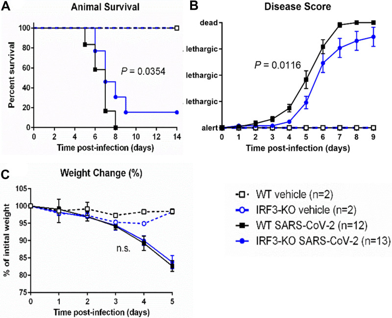
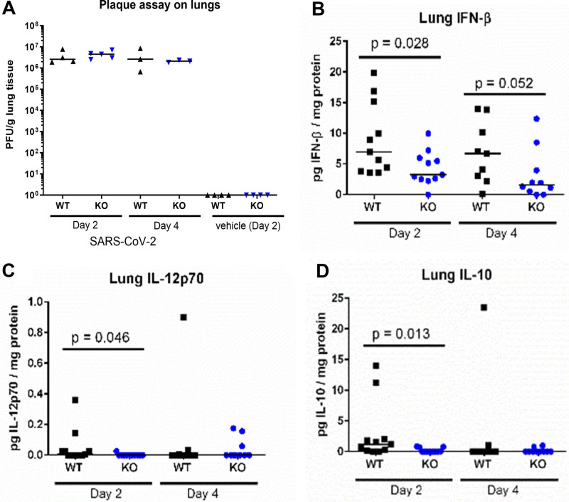
Results: Upon infection with SARS-CoV-2, K18-ACE2 mice showed a dose-dependent disease, characterized by mortality, lethargy, weight loss, and lung pathology, reminiscent of clinical COVID-19. However, K18-ACE2 mice lacking IRF3 were protected from severe disease with reduced mortality (84.6% vs. 100%) and disease score. We found that IRF3 promoted IFN-β production in the lungs and reprogrammed the cytokine profile, while viral load in the lungs was similar in the presence or absence of IRF3.
Interpretations and conclusions: These data indicated that IRF3 played a detrimental role in murine COVID-19 associated with changes in IFN-β and inflammatory cytokines.
Keywords: COVID-19; K18-ACE2 mice; cytokines; human angiotensin-converting enzyme 2 transgenic mice; interferon regulatory factor 3; interferons.
Copyright © 2025 The Authors. Published by Wolters Kluwer Health, Inc. on behalf of the Society of Critical Care Medicine.
Figures
Similar articles
-
Severe Acute Respiratory Syndrome Coronavirus 2 Variant Infection Dynamics and Pathogenesis in Transgenic K18-hACE2 and Inbred Immunocompetent C57BL/6J Mice.Viruses. 2025 Mar 30;17(4):500. doi: 10.3390/v17040500. Viruses. 2025. PMID: 40284943 Free PMC article.
-
CCR2 Signaling Restricts SARS-CoV-2 Infection.mBio. 2021 Dec 21;12(6):e0274921. doi: 10.1128/mBio.02749-21. Epub 2021 Nov 9. mBio. 2021. PMID: 34749524 Free PMC article.
-
Physical interventions to interrupt or reduce the spread of respiratory viruses.Cochrane Database Syst Rev. 2023 Jan 30;1(1):CD006207. doi: 10.1002/14651858.CD006207.pub6. Cochrane Database Syst Rev. 2023. PMID: 36715243 Free PMC article.
-
Type I Interferon, Induced by Adenovirus or Adenoviral Vector Infection, Regulates the Cytokine Response to Lipopolysaccharide in a Macrophage Type-Specific Manner.J Innate Immun. 2024;16(1):226-247. doi: 10.1159/000538282. Epub 2024 Mar 25. J Innate Immun. 2024. PMID: 38527452 Free PMC article.
-
Antibody tests for identification of current and past infection with SARS-CoV-2.Cochrane Database Syst Rev. 2022 Nov 17;11(11):CD013652. doi: 10.1002/14651858.CD013652.pub2. Cochrane Database Syst Rev. 2022. PMID: 36394900 Free PMC article.
References
MeSH terms
Substances
Grants and funding
LinkOut - more resources
Full Text Sources
Medical
Miscellaneous
