

Automated program using convolutional neural networks for objective and reproducible selection of corneal confocal microscopy images
- PMID: 40103638
- PMCID: PMC11915551
- DOI: 10.1177/20552076251326223
Automated program using convolutional neural networks for objective and reproducible selection of corneal confocal microscopy images
Abstract
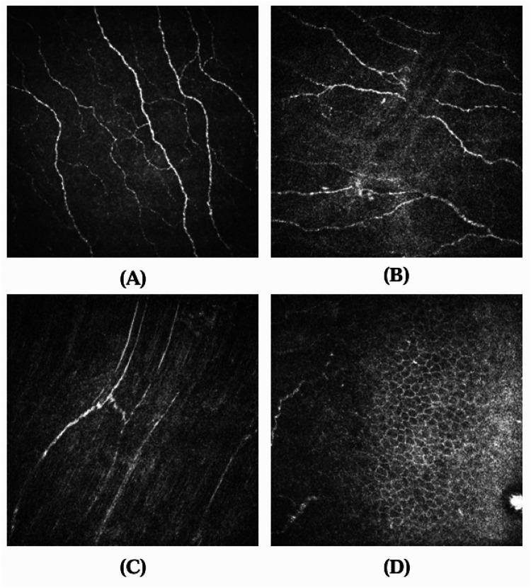
Objective: Diabetic peripheral neuropathy (DPN) is a common complication of diabetes, posing a significant risk for foot ulcers and amputation. Corneal confocal microscopy (CCM) is a rapid, noninvasive method to assess DPN by analysing corneal nerve fibre morphology. However, selecting high-quality representative images remains a critical challenge.
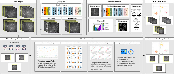
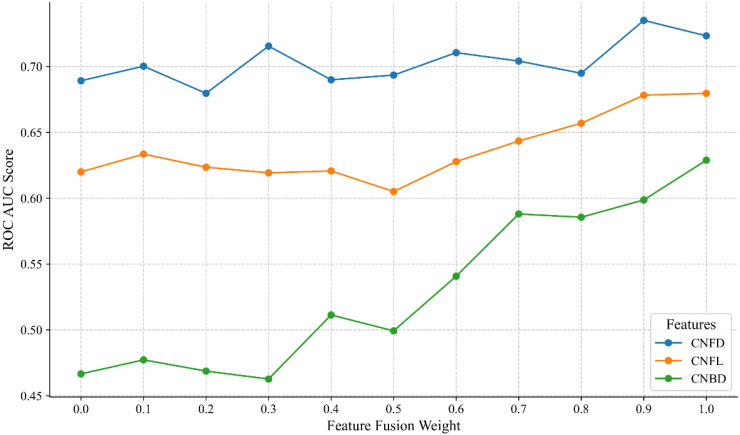
Methods: In this study, we propose a fully automated CCM image-selection algorithm based on deep learning feature extraction using ResNet-18 and unsupervised clustering. The algorithm consistently identifies representative images by balancing non-redundancy and representativeness, ensuring objectivity and reproducibility.
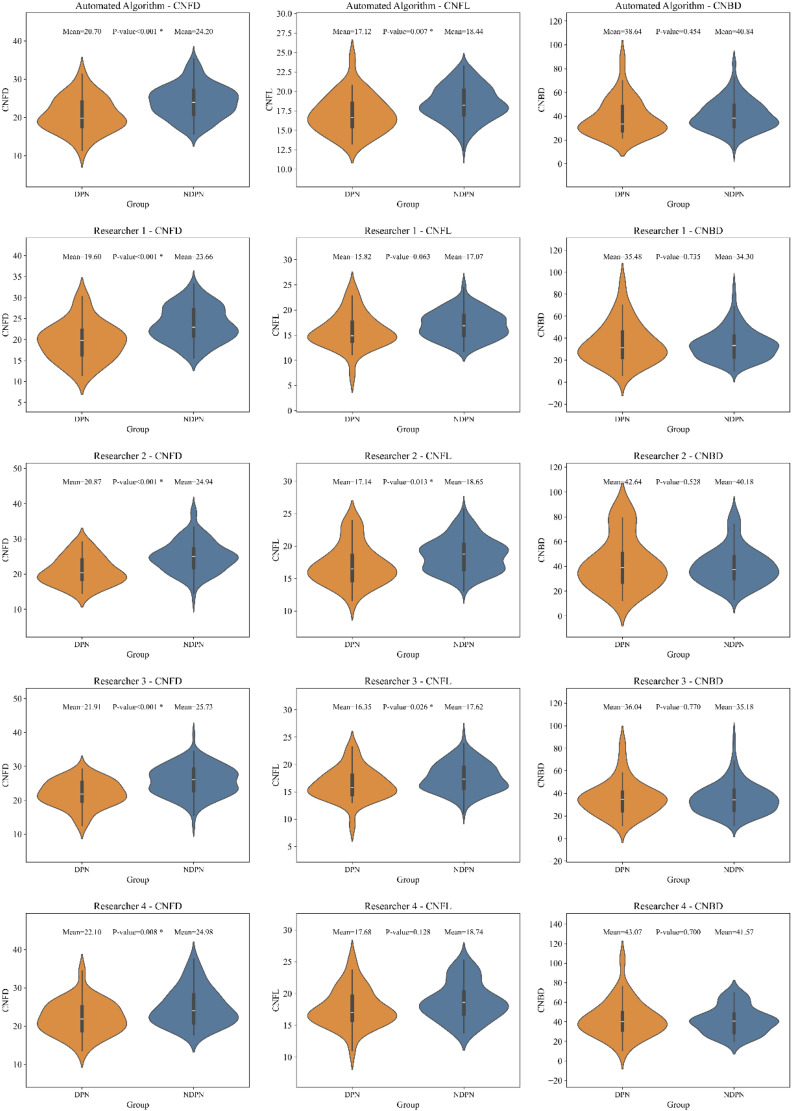
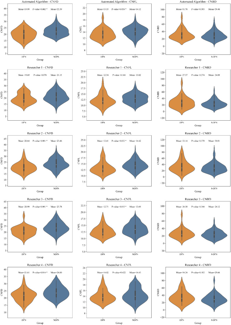
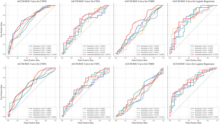
Results: When validated against manual selection by researchers with varying expertise levels, the algorithm demonstrated superior performance in distinguishing DPN and reduced inter-observer variability. It completed the analysis of hundreds of images within 1 s, significantly enhancing diagnostic efficiency. Compared with traditional manual selection, the proposed method achieved higher diagnostic accuracy for key morphological parameters, including corneal nerve fibre density, length, and branch density.
Conclusion: The algorithm is open source and compatible with standard CCM workflows, offering researchers and clinicians a robust and efficient tool for DPN diagnosis. Further, multicentre studies are needed to validate these findings in diverse populations.
Keywords: Deep learning; confocal microscopy; diabetic neuropathy.
© The Author(s) 2025.
Conflict of interest statement
The authors declared no potential conflicts of interest with respect to the research, authorship, and/or publication of this article.
Figures
Similar articles
-
An artificial intelligence-based deep learning algorithm for the diagnosis of diabetic neuropathy using corneal confocal microscopy: a development and validation study.Diabetologia. 2020 Feb;63(2):419-430. doi: 10.1007/s00125-019-05023-4. Epub 2019 Nov 12. Diabetologia. 2020. PMID: 31720728 Free PMC article.
-
Artificial Intelligence and Corneal Confocal Microscopy: The Start of a Beautiful Relationship.J Clin Med. 2022 Oct 20;11(20):6199. doi: 10.3390/jcm11206199. J Clin Med. 2022. PMID: 36294519 Free PMC article. Review.
-
Corneal confocal microscopy for the diagnosis of diabetic peripheral neuropathy: A systematic review and meta-analysis.J Diabetes Investig. 2022 Jan;13(1):134-147. doi: 10.1111/jdi.13643. Epub 2021 Aug 27. J Diabetes Investig. 2022. PMID: 34351711 Free PMC article.
-
Development of a transformer-based deep learning algorithm for diabetic peripheral neuropathy classification using corneal confocal microscopy images.Front Cell Dev Biol. 2024 Oct 14;12:1484329. doi: 10.3389/fcell.2024.1484329. eCollection 2024. Front Cell Dev Biol. 2024. PMID: 39469112 Free PMC article.
-
Utility of corneal confocal microscopy for assessing mild diabetic neuropathy: baseline findings of the LANDMark study.Clin Exp Optom. 2012 May;95(3):348-54. doi: 10.1111/j.1444-0938.2012.00740.x. Epub 2012 Apr 29. Clin Exp Optom. 2012. PMID: 22540156
Cited by
-
ConNeCT: weakly supervised corneal confocal microscopy image inpainting network based on a diffusion model.Biomed Opt Express. 2025 Jun 9;16(7):2615-2630. doi: 10.1364/BOE.562924. eCollection 2025 Jul 1. Biomed Opt Express. 2025. PMID: 40677830 Free PMC article.
References
-
- Feldman EL, Callaghan BC, Pop-Busui R, et al. Diabetic neuropathy. Nat Rev Dis Primers 2019; 5: 41. - PubMed
LinkOut - more resources
Full Text Sources
