. 2025 Mar 13;16(15):6468-6479.
doi: 10.1039/d4sc07994a. eCollection 2025 Apr 9.

Modular access to nucleobase GFP-surrogates: pH-responsive smart probes for ratiometric nucleic acid diagnostics

Affiliations

Modular access to nucleobase GFP-surrogates: pH-responsive smart probes for ratiometric nucleic acid diagnostics

Keenan T Regan et al. Chem Sci. .

Abstract

We have utilized a modular on-strand aldol approach to synthesize chalcone-based fluorescent molecular rotors (FMRs) bearing phenolic oxygen donors that mimic the natural tyrosine (Tyr66) chromophore 4-hydroxybenzylidene-imidazolinone (HBI) within green fluorescent proteins (GFPs). Leveraging the FMRs' propensity to undergo non-radiative decay via twisted intramolecular charge transfer upon excitation within certain microenvironments, we have addressed the longstanding issues of poor brightness (ε max × Φ fl) and weak turn-on responses for GFP-surrogates within nucleic acids. To demonstrate its potential and lay the groundwork for future applications, these FMRs were incorporated into NarI12 and TBA15 oligonucleotides with canonical (A, C, T, G) or locked nucleic acids (LNAs) (TL, AL) as flanking bases. The resulting duplexes and G-quadruplexes (GQs) were studied using fluorescence spectroscopy, molecular dynamics simulations, and quantum mechanical calculations, yielding a comprehensive understanding of their structural and photophysical properties in DNA, DNA : RNA, and GQ contexts. Electron-rich chalcones favor neutral phenol excitation (ROH) to afford both phenol (ROH*) and phenolate (RO-*) emission, with the latter generated through an intermolecular excited-state proton transfer process, while electron-deficient chalcones serve as ratiometric excitation indicators, due to their photoacidity. The surrogates display strong turn-on responses (up to 154-fold) in a GQ → duplex topology switch with flanked LNAs, giving Φ fl up to 0.58 and molar brightness ∼ 15 000 cm-1 M-1 in the duplex. By synergizing the NA sequence and probe, we achieve a switchable ON-to-OFF photoinduced electron transfer, resulting in a 134-fold turn-on emission response to pH. Our findings are the first to optimize the performance of GFP-surrogates as internal nucleobase replacements and suggest multiple ways in which they may be useful tools for NA diagnostics.


Conflict of interest statement

There are no conflicts to declare.

Figures

Fig. 1
Fig. 1. (A) (i) Previously attempted nucleic acid GFP-surrogates, (ii) TICT schematic with and without nucleic acid hybridization, and (iii) light-up responses for previously attempted nucleic acid GFP-surrogates versus this work. (B) This work employs phenolic O-donor aldehydes to generate phenolic-6HI surrogates structurally related to the GFP chromophore HBI.
Fig. 2
Fig. 2. General scheme for the on-strand aldol condensation using either piperidine (1) or morpholine (2), with respective surrogate structures and yields from HPLC analysis.
Fig. 3
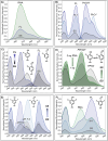
Fig. 3. (A) Emission spectra of PhOH6HI (SS vs. FL DNA duplex vs. DNA : RNA hybrid duplex, green trace) highlighting hybridization impact on relative emission intensity, λex = 390 nm. (B) Excitation/emission spectra of PhOH6HI (SS vs. FL DNA vs. N-1 DNA, blue trace) highlighting hybridization impact on ESPT to afford PhO* emission from PhOH excitation at 380 nm. (C) Excitation/emission spectra of FPhOH6HI (SS vs. FL DNA) highlighting ratiometric excitation response to hybridization at pH 7.4, excitation spectra recorded with λem = 570 nm. (D) Excitation spectra (λem = 570 nm) for FPhOH6HI displaying phenolate (FPhO) excitation in the SS (black trace) to predominately phenol (FPhOH) excitation in the DNA : RNA hybrid duplex (green traces) at pH 7.4. (E) Excitation/emission spectra of DFPhOH6HI (SS vs. FL DNA at pH 7.4 and 6.0) highlighting ratiometric excitation response to hybridization at pH 6.0. (F) Hybridization impact on excitation spectra for DFPhOH6HI at pH 7.4 displaying phenolate (DFPhO) excitation in the SS (black trace) to progressive increases in phenol (DFPhOH) excitation upon hybridization to afford the N-1 DNA (blue trace), FL DNA (purple trace) and DNA : RNA hybrid duplex (green trace).
Fig. 4
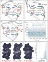
Fig. 4. Representative MD structures of Narl12 DS containing PhOH6HI (white), images are side views from the major groove with water, ions, and nonpolar hydrogen atoms hidden for clarity. (A) PhOH6HI opposite dC18 (pink) in FL DNA, (B) PhOH6HI opposite N-1 strand, and (C) PhOH6HI opposite C18 (pink) in DNA : RNA hybrid. (D) Average co-planar angle and number of 90° rotations for FL, N-1, and RNA : DNA systems. A 100 ns moving average is superimposed onto the raw data to highlight trends. (E) MD-determined solvent accessible surface areas for (i) PhOH6HI in FL DNA, (ii) PhOH6HI in N-1 DNA, and (iii) PhOH6HI in DNA : RNA hybrid, and the percent shielding of the phenolic oxygen atom.
Fig. 5
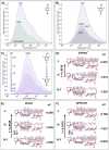
Fig. 5. (A) Emission spectra of PhOH6HI in the FL NarI12 DNA duplex, highlighting the effect of flanking sequence on relative emission intensity (excitation maxima 390 nm). (B) Emission spectra of DFPhOH6HI in the FL NarI12 DNA duplex, highlighting the effect of flanking sequence on relative emission intensity (excitation maxima 450 nm). (C) Emission spectra of DFPhOH6HI in GXG NarI12 as a function of pH (5.5–8.0, excitation maxima 450 nm). (D–F) Minimum energy structures for DFPhO6HI (D), PhOH6HI (E), and DFPhOH6HI (F), flanked by dG (purple), as evaluated at the SMD-ωB97X-D/Def2-TZVP//SMD-ωB97X-D/Def2-SVP (water) level of theory with their HOMO − 1, HOMO, and LUMO (H − 1, H, L) energy levels (isovalue = 0.02). The oscillator strength f (S0 → S1) was determined through a TD-DFT calculation at the SMD-ωB97X-D/Def2-TZVP (water) level of theory.
Fig. 6
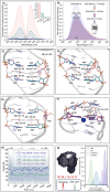
Fig. 6. (A) Excitation/emission spectra for DFPhOH6HI (pink trace), FPhOH6HI (teal trace), DMePhOH6HI (black trace), and PhOH6HI (blue trace) at G8 of TBA15 recorded in Tris–HCl pH 8.0, 50 mM KCl, 50 mM MgCl2. Irel given in the inset. (B) Fluorescence emission spectra highlighting turn-on responses to hybridization by PhOH6HI at the G8-site of TBA15 in the GQ vs. DNA FL duplex (red trace), DNA : RNA hybrid (blue trace), and DNA : RNA–AL hybrid (purple trace) in Tris–HCl pH 8.0, 50 mM KCl and 50 mM MgCl2. Representative MD structures of TBA15 containing PhOH6HI (white) in (C) DNA : RNA hybrid, (D) PhOHDNA : RNA–AL hybrid, (E) DNA : RNA–TL hybrid, and (F) BA15 GQ K+. Water, ions, and nonpolar hydrogen atoms are hidden for clarity. (G) Average co-planar angle and number of 90° rotations for TBA15 GQ K+, TBA DNA : RNA–AT, TBA DNA : RNA–AL, and TBA RNA : DNA. A 100 ns moving average is superimposed on the raw data to highlight trends. (H) MD-determined solvent accessible surface area for PhOH6HI (white) for TBA GQ K+, and percent solvent shield occupancy for the phenolic oxygen.
Fig. 7
Fig. 7. Performance of GFP-surrogates versus other FMR and smart surrogates used for NA diagnostics.

Similar articles

References

    1. Day R. N. Davidson M. W. The Fluorescent Protein Palette: Tools for Cellular Imaging. Chem. Soc. Rev. 2009;38:2887–2921. doi: 10.1039/B901966A. - DOI - PMC - PubMed
    1. Hoffman R. M. The Multiple Uses of Fluorescent Proteins to Visualize Cancer in Vivo. Nat. Rev. Cancer. 2005;5:796–806. doi: 10.1038/nrc1717. - DOI - PubMed
    1. Newman R. H. Fosbrink M. D. Zhang J. Genetically Encodable Fluorescent Biosensors for Tracking Signaling Dynamics in Living Cells. Chem. Rev. 2011;111:3614–3666. doi: 10.1021/cr100002u. - DOI - PMC - PubMed
    1. Tsien R. Y. Constructing and Exploiting the Fluorescent Protein Paintbox (Nobel Lecture) Angew. Chem., Int. Ed. 2009;48:5612–5626. doi: 10.1002/anie.200901916. - DOI - PubMed
    1. Steffen F. D. Khier M. Kowerko D. Cunha R. A. Börner R. Sigel R. K. O. Metal Ions and Sugar Puckering Balance Single-Molecule Kinetic Heterogeneity in RNA and DNA Tertiary Contacts. Nat. Commun. 2020;11:104. doi: 10.1038/s41467-019-13683-4. - DOI - PMC - PubMed