. 2025 Mar 4:19:1558605.
doi: 10.3389/fncel.2025.1558605. eCollection 2025.

Morphology and connectivity of retinal horizontal cells in two avian species

Affiliations

Morphology and connectivity of retinal horizontal cells in two avian species

Anja Günther et al. Front Cell Neurosci. .

Abstract

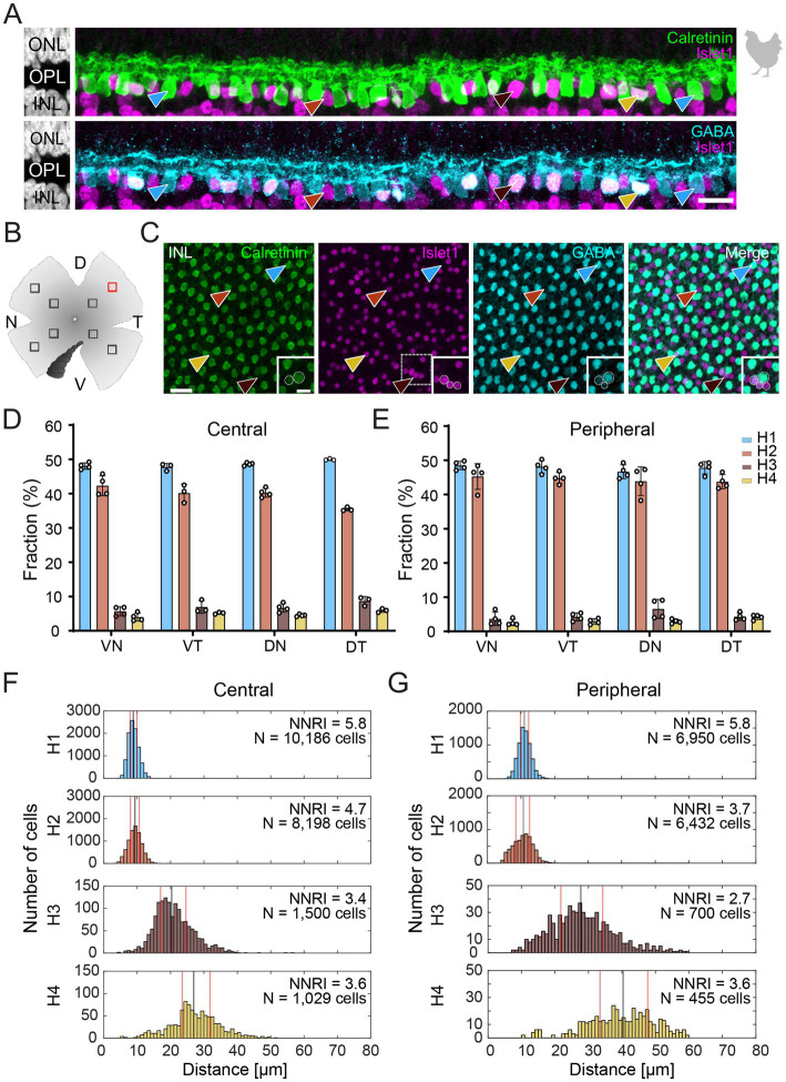
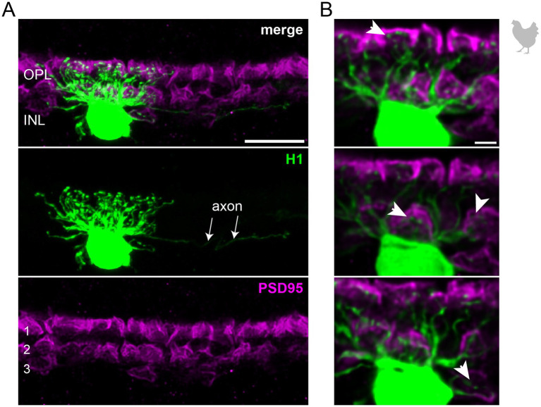
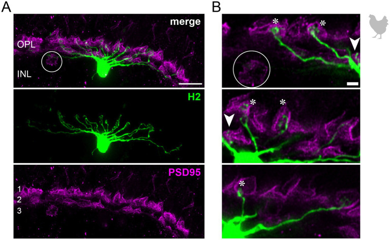
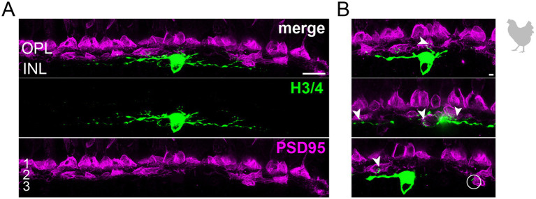
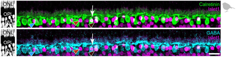
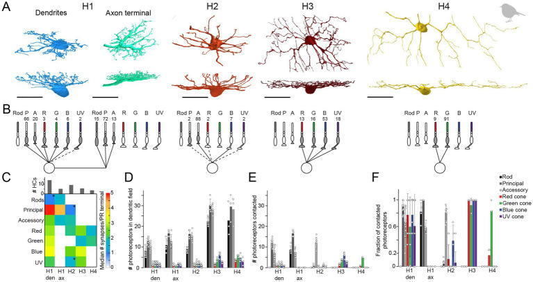
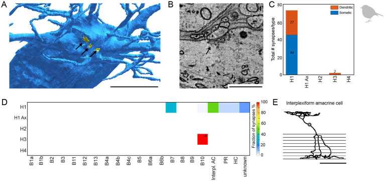
In the outer vertebrate retina, the visual signal is separated into intensity and wavelength information. In birds, seven types of photoreceptors (one rod, four single cones, and two members of the double cone) mediate signals to >20 types of second-order neurons, the bipolar cells and horizontal cells. Horizontal cells contribute to color and contrast processing by providing feedback signals to photoreceptors and feedforward signals to bipolar cells. In fish, reptiles, and amphibians they either encode intensity or show color-opponent responses. Yet, for the bird retina, the number of horizontal cell types is not fully resolved and even more importantly, the synapses between photoreceptors and horizontal cells have never been quantified for any bird species. With a combination of light microscopy and serial EM reconstructions, we found four different types of horizontal cells in two distantly related species, the domestic chicken and the European robin. In agreement with some earlier studies, we confirmed two highly abundant cell types (H1, H2) and two rare cell types (H3, H4), of which H1 is an axon-bearing cell, whereas H2-H4 are axonless. H1 cells made chemical synapses with one type of bipolar cell and an interplexiform amacrine cell at their soma. Dendritic contacts of H1-H4 cells to photoreceptors were type-specific and similar to the turtle retina, which confirms the high degree of evolutionary conservation in the vertebrate outer retina. Our data further suggests that H1 and potentially H2 cells may encode intensity, whereas H3 and H4 may represent color opponent horizontal cells which may contribute to the birds' superb color and/or high acuity vision.

Keywords: European robin; avian retina; chicken; connectomics; horizontal cells; magnetoreception; photoreceptors; vision.


Conflict of interest statement

The authors declare that the research was conducted without any commercial or financial relationships that could be construed as a potential conflict of interest. The author(s) declared that they were an editorial board member of Frontiers, at the time of submission. This had no impact on the peer review process and the final decision.

Figures

Figure 1
Figure 1
Triple labeling with Calretinin, GABA, and Islet1 allows the differentiation of four types of horizontal cells in the chicken retina. (A) Maximum projections of vertical slices of the chicken retina labeled for Calretinin (Calret), GABA, and Islet1. Based on these markers, four different types of horizontal cells can be distinguished: H1: Calret+/GABA+ (blue arrowhead); H2: Islet1+ (red arrowhead); H3: Islet+/GABA+ (brown arrowhead), and H4: Calret+/GABA+/Islet1+ (yellow arrowhead). (B) Schematic of the flat-mounted retina of a chicken with cuts in the nasal (N), dorsal (D), temporal (T) and ventral (V) side to flatten the tissue. The dark comb-like structure represents the pecten. Four image stacks each were taken from the central and peripheral retina, marked by the squares (not to scale). An example from the dorsal-temporal region (red square) is shown in (C). (C) Flat-mounted retina from the chicken retina, labeled for Calret, GABA and Islet1. Focus is on the INL. Arrowheads point to the different types of horizontal cells with the same color code as in (B). The area marked with a dashed square is shown enlarged in the insets for all three channels; this area contains all four horizontal cell types. Dashed circles represent the cell bodies labeled by the respective marker. (D,E) Bar graphs representing the fraction of horizontal cells per type per retinal area. Data is given as mean ± standard deviation; n = 4 retinas from 3 animals, except for VT and DT in (D) where n = 3 retinas from 2 animals. (F,G) Distribution of nearest neighbor distances for each of the four horizontal cell types in the central (F) and peripheral chicken retina (G). The black and red vertical lines spanning the entire box give the median and the lower and upper quartiles of the nearest neighbor distances, respectively. NNRI represents the ratio between the mean of the nearest neighbor distance and the standard deviation of the cellular array (NNRI = nearest neighbor regularity index). Values above 1.9 are considered regular (Cook, 1996). Scale bars: (A) 10 μm; (C) 20 μm; inset: 10 μm.
Figure 2
Figure 2
The axon-bearing H1 horizontal cell in the peripheral chicken retina contacts all cones. (A) Maximum projection of a dye-injected H1 horizontal cell, revealing a dense and narrow dendritic field and an axon (arrows). Double labeling with PSD95 reveals the stratification of photoreceptor terminals in three layers (labeled 1–3) of the outer plexiform layer (OPL). Please note that for PSD95, the maximum projection of a substack is shown to better illustrate the three layers. (B) Single optical sections at different positions of the confocal stack. H1 cell dendrites contact cones in all three layers of the OPL (arrowheads). Whether or not rods are also contacted by the dendritic field could not be resolved. Please note that the H1 soma was saturated on purpose to better reveal the thin dendrites. Scale: 10 μm (A), 2 μm (B).
Figure 3
Figure 3
H2 horizontal cells in the peripheral chicken retina make selective contacts to the accessory member of the double cone photoreceptors. (A) Dye-injected H2 horizontal cell, revealing a large dendritic tree which is only branching in the terminal endings. A maximum projection is shown. Double labeling with PSD95 reveals the stratification of photoreceptor terminals in three layers (labeled 1–3) of the outer plexiform layer (OPL). Please note that for PSD95, the maximum projection of a substack is shown to better illustrate the three layers. (B) Maximum projections of substacks (12–18 optical sections). Please note that the H2 soma was saturated on purpose to better reveal the thin dendrites. The vast majority of H2 cell dendrites contact the accessory members of the double cones in the most distal OPL layer (1, asterisks), while avoiding photoreceptor terminals in layer 3 which belong to violet and blue cones (Balaji et al., 2023; Mariani, 1987), marked by a circle. However, one dendrite seems to contact a green or red cone in layer 2 of the OPL (arrowhead). Scale: 10 μm (A), 2 μm (B).
Figure 4
Figure 4
Wide-field horizontal cell (H3 or H4) in the peripheral chicken retina avoiding the outermost layer of the outer plexiform layer. (A) Maximum projection of a dye-injected wide-field horizontal cell, revealing a flat dendritic field with processes mostly confined to layer 2 of the outer plexiform layer. Whether this cell represents an H3 or H4 cell is not possible to discern. Double labeling with PSD95 reveals the stratification of photoreceptor terminals in three layers (labeled 1–3) of the outer plexiform layer (OPL). Please note that for PSD95, the maximum projection of a substack is shown to better illustrate the three layers. (B) Maximum projections of substacks (30–35 optical sections) of the confocal stack. H3/4 cell dendrites contact mostly red and/or green cones in layer 2 of the OPL (arrowheads), while avoiding rods and double cones in the most distal layer (1) of the OPL. Rarely, H3/4 cell dendrites reach out to violet/blue cones (circle) in the most proximal layer (3) of the OPL (Balaji et al., 2023; Mariani, 1987). However, whether these dendrites which do not invaginate the cone pedicle represent true contacts is unclear. Please note that the H3/4 soma was saturated on purpose to better reveal the thin dendrites. Scale: 10 μm (A), 2 μm (B).
Figure 5
Figure 5
Triple labeling with Calretinin, GABA, and Islet1 allows to differentiate only three types of horizontal cells in the European robin retina. Maximum projections of vertical slices of the European robin retina labeled for Calretinin (Calret), GABA, and Islet1. Based on these markers, three different types of horizontal cells were distinguished: H1: Calret+/GABA+ (blue arrowhead); H2: Islet1+ (red arrowhead); H3: Islet+/GABA+ (brown arrowhead). The white arrow points to a cell which seems to express all three markers, but inspection of the confocal stack showed that an Islet1+ H2 cell lies behind a Calret+/GABA+ H1 cell. Scale bar: 10 μm.
Figure 6
Figure 6
Quantification of horizontal cell type specific contacts to photoreceptors. (A) Volume reconstructions of each horizontal cell type. In total, we identified four horizontal cell types namely H1 (dendrites n = 35, axons n = 5), H2 (n = 21), H3 (n = 6), and H4 (n = 3). All scale bars: 20 μm. (B) Network motifs for each horizontal cell type. The numbers above each photoreceptor type represent the fraction of synapses/photoreceptor type in percent. Dashed lines indicate that this photoreceptor cell type was rarely (<3%) contacted by the respective horizontal cell. P, principal member of a double cone; A, accessory member of a double cone; R, red single cone; G, green single cone; B, blue single cone; UV, ultraviolet single cone. (C) Connectivity matrix with the median number of synapses/photoreceptor (PR) terminal per horizontal cell type. Histograms above the connectivity matrix indicate the number of cells that were included in the analysis. Connections between horizontal cells and photoreceptor cells that are highlighted by an asterisk only occurred in a few individual horizontal cells; den = dendrites; ax = axon terminal. (D) Mean number ± standard deviation (SD) of rod and cone terminals within the dendritic field of different horizontal cells. (E) Mean number ± SD of contacted photoreceptor cells/type for different horizontal cell types. (F) Mean fraction ± SD of contacted photoreceptors from photoreceptors in the dendritic field of different horizontal cell types.
Figure 7
Figure 7
H1 horizontal cells form conventional synapses at their dendrites and somata to specific bipolar and interplexiform amacrine cells. (A) Volume reconstructed soma of an H1 horizontal cell (blue) with arrows pointing to the locations of conventional synapses highlighted in yellow. Scale bar: 5 μm. (B) Electron microscopic image of a conventional synapse at the soma of an H1 cell. The image originates from the previously published mSEM dataset (Günther et al., 2024). Scale bar: 1 μm. (C) Total number of conventional synapses for each HC type separated by their location either on the cell body or the dendrites. In total, we inspected 21 H1, 18 H2, 6 H3 and 3 H4 horizontal cells. (D) Connectivity matrix with fractionated number of synapses for each postsynaptic cell type in percent. Only two small synapses were found at H3 horizontal cell dendrites (marked with *). Postsynaptic cells from the H3 horizontal cell synapses resembled the previously described B10 bipolar cell in the chicken (Günther et al., 2021). Ax = axon terminal; B1-B10, bipolar cell types; interpl. AC, interplexiform amacrine cell; HC, horizontal cell; PR, photoreceptor. (E) Skeletonized postsynaptic interplexiform amacrine cell, stratifying in the outer and inner plexiform layer. Scale bar: 50 μm.

Similar articles

Cited by

  • A standardized nomenclature for the rods and cones of the vertebrate retina.
    Baden T, Angueyra JM, Bosten JM, Collin SP, Conway BR, Cortesi F, Dedek K, Euler T, Novales Flamarique I, Franklin A, Haverkamp S, Kelber A, Neuhauss SCF, Li W, Lucas RJ, Osorio DC, Shekhar K, Tommasini D, Yoshimatsu T, Corbo JC. Baden T, et al. PLoS Biol. 2025 May 7;23(5):e3003157. doi: 10.1371/journal.pbio.3003157. eCollection 2025 May. PLoS Biol. 2025. PMID: 40333813 Free PMC article.
  • AAV-mediated transduction of songbird retina.
    Seth PK, Heyers D, Satish B, Mendoza E, Haase K, Borowsky L, Musielak I, Koch KW, Feederle R, Scharff C, Dedek K, Mouritsen H. Seth PK, et al. Front Physiol. 2025 Mar 19;16:1549585. doi: 10.3389/fphys.2025.1549585. eCollection 2025. Front Physiol. 2025. PMID: 40177359 Free PMC article.

References

    1. Ammermüller J., Kolb H. (1996). Functional architecture of the turtle retina. Prog. Retin. Eye Res. 15, 393–433. doi: 10.1016/1350-9462(96)00009-2 - DOI
    1. Ammermüller J., Muller J. F., Kolb H. (1995). The organization of the turtle inner retina. II. Analysis of color-coded and directionally selective cells. J. Comp. Neurol. 358, 35–62. doi: 10.1002/cne.903580103, PMID: - DOI - PubMed
    1. Araki M., Kimura H. (1991). GABA-like immunoreactivity in the developing chick retina: differentiation of GABAergic horizontal cell and its possible contacts with photoreceptors. J. Neurocytol. 20, 345–355. doi: 10.1007/BF01355531, PMID: - DOI - PubMed
    1. Asi H., Perlman I. (1998). Neural interactions between cone photoreceptors and horizontal cells in the turtle (Mauremys caspicd) retina. Vis. Neurosci. 15, 1–13. doi: 10.1017/s0952523898146047, PMID: - DOI - PubMed
    1. Baden T. (2024). The vertebrate retina: a window into the evolution of computation in the brain. Curr. Opin. Behav. Sci. 57:101391. doi: 10.1016/j.cobeha.2024.101391, PMID: - DOI - PMC - PubMed

LinkOut - more resources