Case Reports
. 2025 Feb 26;9(3):ytaf109.
doi: 10.1093/ehjcr/ytaf109. eCollection 2025 Mar.

Haemodynamic spectrum in heritable pulmonary arterial hypertension: a continuum from pre-capillary to combined pulmonary hypertension-case series

Affiliations
Case Reports

Haemodynamic spectrum in heritable pulmonary arterial hypertension: a continuum from pre-capillary to combined pulmonary hypertension-case series

Irene Martín de Miguel et al. Eur Heart J Case Rep. .

Abstract

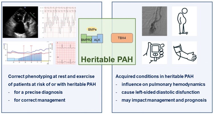
Background: Heritable pulmonary arterial hypertension (PAH) is a rare form of pre-capillary pulmonary hypertension that typically affects young patients. With increased survival and subsequent ageing of these patients, newly acquired cardiovascular conditions may influence the pulmonary haemodynamic profile and impact management.

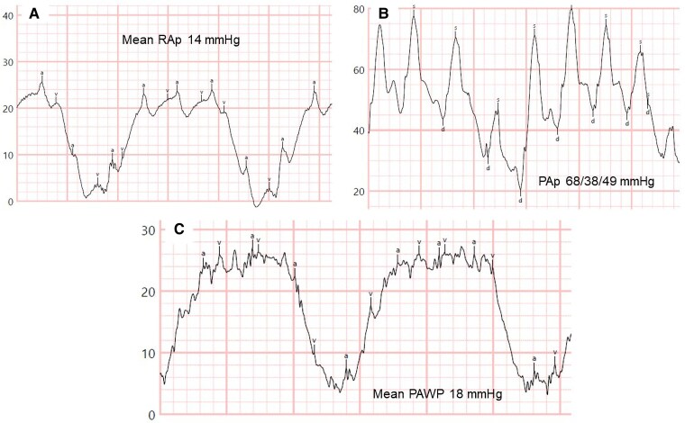
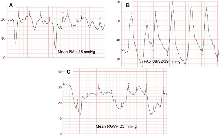
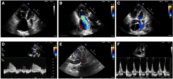
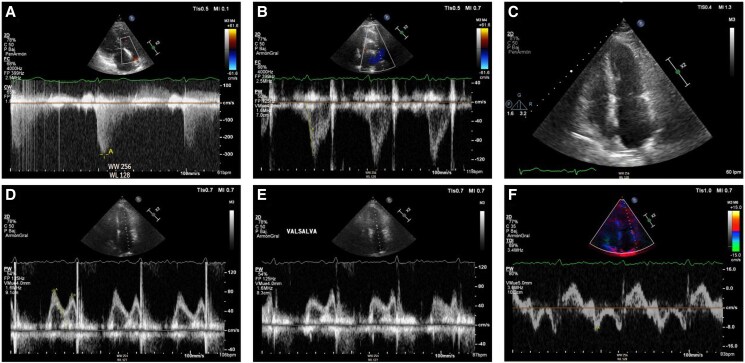
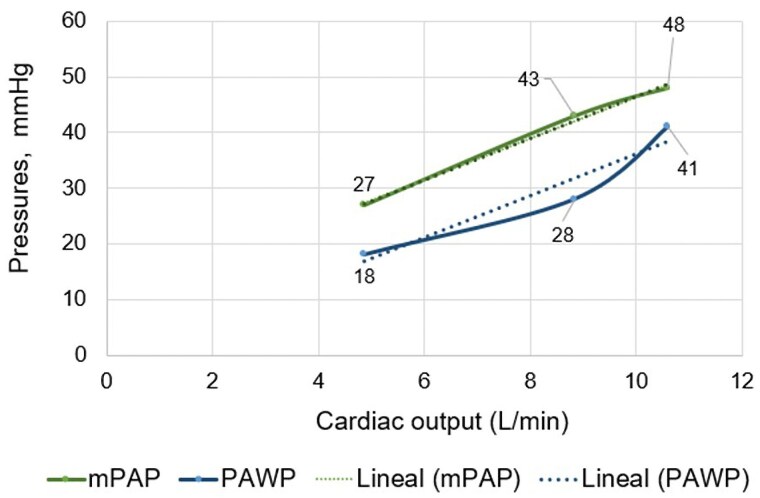
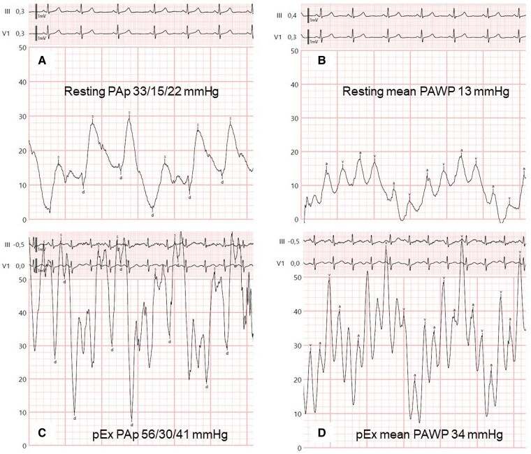
Case summary: We report a case series of four patients with mutations in genes associated with PAH to illustrate the spectrum of pulmonary haemodynamics under the influence of superimposed acquired conditions. The first two cases involve patients with a long-standing diagnosis of heritable PAH and severe pre-capillary pulmonary hypertension, who developed overt left-sided diastolic dysfunction later in follow-up due to the acquisition of multiple cardiovascular comorbidities. The second two cases describe patients with a genetic pre-disposition to develop PAH and conditions that are risk factors for left heart disease, with mild elevation of resting pulmonary pressures, in whom exercise right heart catheterization unmasked occult left-sided diastolic dysfunction.

Discussion: Pulmonary haemodynamics are complex and dynamic over time, even in patients with or at risk of heritable PAH, when additional acquired cardiovascular conditions emerge. Correct phenotyping at diagnosis and during follow-up of patients at risk of heritable PAH, along with a clear understanding of the underlying pulmonary haemodynamic profile, is crucial for appropriate management.

Keywords: Cardiovascular comorbidities; Case series; Diastolic dysfunction; Exercise right heart catheterization; Heritable pulmonary arterial hypertension; Post-capillary pulmonary hypertension; Pre-capillary pulmonary hypertension.


Conflict of interest statement

Conflict of interest. None declared.

Figures

Figure 1
Figure 1
Resting right heart catheterization tracings during follow-up of Case 1 at time of hospital admission. (A) Mean right atrial pressure 14 mmHg. (B) Pulmonary artery pressures 68/38/49 mmHg. (C) Mean pulmonary arterial wedge pressure 18 mmHg. Note the significant respiratory variations due to obesity. PAp, pulmonary artery pressures; PAWP, pulmonary arterial wedge pressure; RAp, right atrial pressure.
Figure 2
Figure 2
Resting right heart catheterization tracings during follow-up of Case 2 at time of hospital admission. (A) Mean right atrial pressure 18 mmHg. (B) Pulmonary artery pressures 68/32/39 mmHg. (C) Mean pulmonary arterial wedge pressure 23 mmHg. PAp, pulmonary artery pressures; PAWP, pulmonary arterial wedge pressure; RAp, right atrial pressure.
Figure 3
Figure 3
Transthoracic echocardiography during follow-up of Case 2 at time of hospital admission. (A) Apical four chamber view depicting a severely dilated right ventricle with the following dimensions: end-diastolic right ventricular basal diameter 52 mm, end-diastolic right ventricular mid diameter 55 mm, end-diastolic right ventricular longitudinal diameter 85 mm, right ventricular end-diastolic area 41.5 cm2. Right ventricular systolic dysfunction was severely reduced (fractional area change 19%, tricuspid annular plane systolic excursion 12 mm with severe tricuspid regurgitation, pulsed Doppler S wave of the tricuspid annulus 6.9 cm/s). Severe right atrial dilation (33 cm2). (B) Right ventricular-focused apical four chamber view showing severe functional tricuspid regurgitation. (C) Apical four chamber view depicting moderate mitral regurgitation due to anterior leaflet prolapse. (D) Moderate functional pulmonary regurgitation allows for estimation of a mean pulmonary artery pressure of 45 mmHg. (E) Dilated inferior vena cava of 24 mm without respiratory variation, estimated right atrial pressure of 15 mmHg. (F) Mitral inflow shows pseudonormal left ventricular filling pattern consistent with Grade 2 diastolic dysfunction (elevated left ventricular filling pressures). Mitral E-wave was 109 cm/s; mitral A-wave was 102 cm/s. Valsalva manoeuvre increased A-wave velocity. Lateral E/e′ ratio was 12.
Figure 4
Figure 4
Transthoracic echocardiography of Case 3 at screening for pulmonary hypertension. (A) Systolic peak tricuspid regurgitation velocity 3.0 m/s. (B) Shortened right ventricular outflow tract acceleration time of pulmonary ejection of 70 ms without evident mid-systolic notch. (C) Apical four chamber view depicting normal right ventricular, right atrial, and left ventricular size and mild left atrial dilation (35 mL/m2). (D and E) Mitral inflow shows pseudonormal left ventricular filling pattern consistent with Grade 2 diastolic dysfunction (elevated left ventricular filling pressures). Mitral E-wave was 82 cm/s; mitral A-wave was 70 cm/s. Valsalva manoeuvre increased A-wave velocity. (F) Reduced lateral mitral e′-wave in tissue Doppler imaging of 8.8 cm/s. Average E/e′ ratio was 11.
Figure 5
Figure 5
Mean pulmonary artery pressure and mean pulmonary arterial wedge pressure/cardiac output slopes at peak exercise of Case 3 during screening for pulmonary hypertension. The mean pulmonary artery pressure/cardiac output slope was 3.7 mmHg/L/min and the mean pulmonary arterial wedge pressure/cardiac output slope was 3.8 mmHg/L/min. CO, cardiac output; mPAP, mean pulmonary artery pressure; PAWP, mean pulmonary arterial wedge pressure.
Figure 6
Figure 6
Pulmonary angiography images of the pulmonary arteriovenous fistulae during embolization. (A) Pulmonary arteriovenous fistula in the basal region of de middle lobe (arrow). (B) Pulmonary arteriovenous fistula in the right lower lobe (arrows). (C) Status post-embolization of the fistula in the basal region of the middle lobe with a 6 mm large Amplatzer device (orange arrow) and of the fistula in the right lower lobe with four 5 mm coils (yellow arrow). Note that three smaller fistulae could technically not be embolized.
Figure 7
Figure 7
Wasserman curves of the cardiopulmonary exercise test of Case 4 during screening for pulmonary hypertension. Exercise was performed on upright cycle ergometer. The test was maximal (respiratory exchange ratio at peak exercise 1.12, Curve 8). Functional capacity was mildly reduced [peak oxygen consumption 16.5 mL/Kg/min (69% of predicted), Curve 3]. There was moderate ventilatory inefficiency: ventilatory equivalent of carbon dioxide at first ventilatory threshold was 38 (138% of predicted) and ventilatory equivalent of oxygen at peak exercise was 46 (Curve 6). Ventilation/carbon dioxide production slope was 34.8 (Curve 4). End-tidal carbon dioxide partial pressure at rest was 24 mmHg, at first ventilatory threshold 30 mmHg, at peak exercise 28 mmHg (Curve 9). AT, first ventilatory threshold; Bf, breathing frequency; BR, breathing reserve; EqCO2, ventilatory equivalent of carbon dioxide; EqO2, ventilatory equivalent of oxygen; HR, heart rate; PETCO2, end-tidal carbon dioxide partial pressure; PETO2, end-tidal oxygen partial pressure; RCP, second ventilatory threshold; RER, respiratory exchange ratio; VE, ventilation; VE/VCO2, ventilation/carbon dioxide production; VO2, oxygen consumption; Vt, tidal volume.
Figure 8
Figure 8
Resting and exercise right heart catheterization tracings of Case 4 during screening for pulmonary hypertension. (A) Resting pulmonary artery pressures 33/15/22 mmHg. (B) Resting mean pulmonary arterial wedge pressure 13 mmHg. (C) Pulmonary artery pressures 56/30/41 mmHg at peak exercise. (D) Mean pulmonary arterial wedge pressure at peak exercise 34 mmHg. PAp, pulmonary artery pressures; PAWP, pulmonary arterial wedge pressure; pEx, peak exercise.
None

Similar articles

References

    1. Hassoun PM. Pulmonary arterial hypertension. N Engl J Med 2021;385:2361–2376. - PubMed
    1. Humbert M, Kovacs G, Hoeper MM, Badagliacca R, Berger RMF, Brida M, et al. 2022 ESC/ERS guidelines for the diagnosis and treatment of pulmonary hypertension: developed by the task force for the diagnosis and treatment of pulmonary hypertension of the European Society of Cardiology (ESC) and the European Respiratory Society (ERS). Endorsed by the International Society for heart and lung transplantation (ISHLT) and the European reference network on rare respiratory diseases (ERN-LUNG). Eur Heart J 2022;43:3618–3731. - PubMed
    1. Martin de Miguel I, Cruz-Utrilla A, Oliver E, Escribano-Subias P. Novel molecular mechanisms involved in the medical treatment of pulmonary arterial hypertension. Int J Mol Sci 2023;24:4147. - PMC - PubMed
    1. Morrell NW, Aldred MA, Chung WK, Elliott CG, Nichols WC, Soubrier F, et al. Genetics and genomics of pulmonary arterial hypertension. Eur Respir J 2019;53:1801899. - PMC - PubMed
    1. Sztrymf B, Coulet F, Girerd B, Yaici A, Jais X, Sitbon O, et al. Clinical outcomes of pulmonary arterial hypertension in carriers of BMPR2 mutation. Am J Respir Crit Care Med 2008;177:1377–1383. - PubMed

Publication types

LinkOut - more resources