Review
. 2025 Mar 4:16:1548535.
doi: 10.3389/fimmu.2025.1548535. eCollection 2025.

Tumor-derived extracellular vesicles: key drivers of immunomodulation in breast cancer

Affiliations
Review

Tumor-derived extracellular vesicles: key drivers of immunomodulation in breast cancer

Jieming Li et al. Front Immunol. .

Abstract

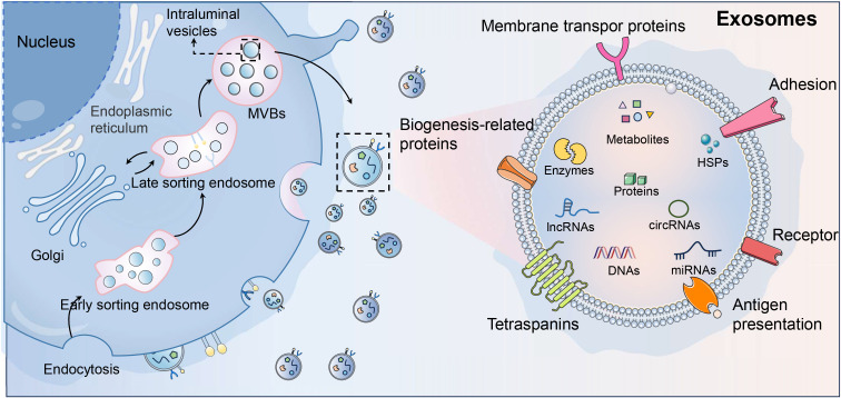
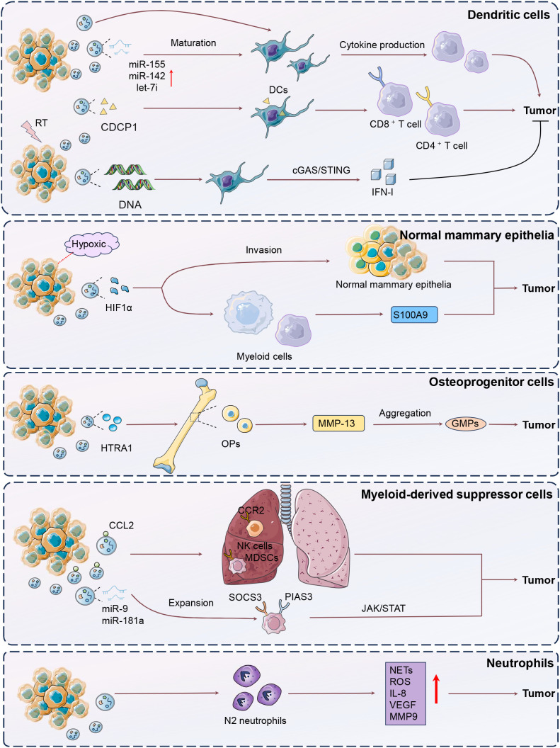
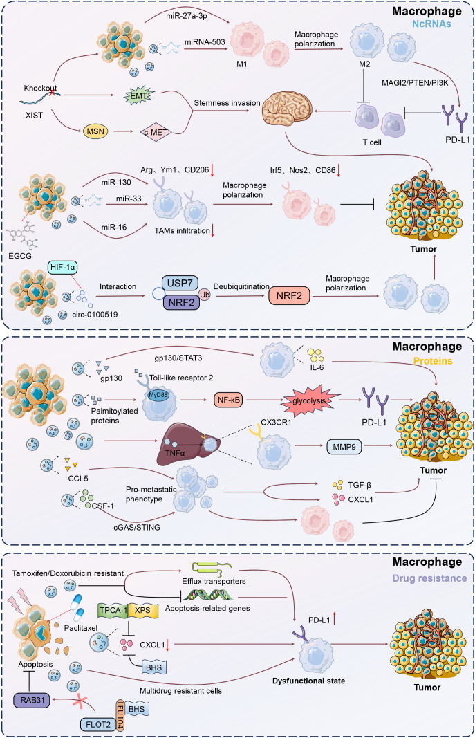
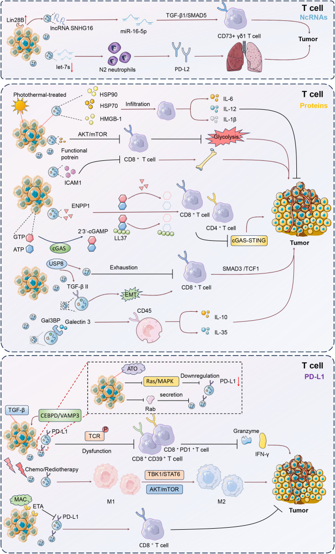
Breast cancer (BC) remains a significant global health challenge characterized by its heterogeneity and treatment complexities. Extracellular vesicles (EVs) are small membranous particles released by cells, facilitating intercellular communication by transporting bioactive molecules such as proteins, lipids, and nucleic acids. Tumor-derived EVs have emerged as pivotal regulators in the tumor microenvironment (TME) and drivers of BC progression. These EVs carry diverse cargoes of bioactive molecules, influencing critical processes such as immune modulation, angiogenesis, and metastasis. By altering the behaviors of immune cells including macrophages, dendritic cells, and T cells, tumor-derived EVs contribute to immune evasion and tumor growth. Furthermore, Tumor-derived EVs play a role in mediating drug resistance, impacting the effectiveness of therapeutic interventions. Understanding the multifaceted roles of BC tumor-derived EVs is essential for the development of innovative therapeutic strategies. Targeting pathways mediated by EVs holds promise for enhancing the efficacy of cancer treatments and improving patient outcomes. This comprehensive review provides insights into the intricate interactions of tumor-derived EVs in immune modulation and BC progression, highlighting potential therapeutic targets and avenues for novel cancer therapies.

Keywords: T cells; breast cancer; extracellular vesicles; immune regulation; macrophages.


Conflict of interest statement

The authors declare that the research was conducted in the absence of any commercial or financial relationships that could be construed as a potential conflict of interest.

Figures

Figure 1
Figure 1
Exosome biogenesis and signature. As the main subtype of extracellular vesicles (EVs), exosomes are key players in intercellular communication within the tumor microenvironment (TME), and originate from the inward budding of the plasma membrane, forming early endosomes that mature into multivesicular bodies (MVBs). The ESCRT machinery and ESCRT-independent pathways involving lipid rafts and tetraspanins (CD63, CD81, CD9) generate intraluminal vesicles (ILVs). MVBs may fuse with lysosomes for degradation or with the plasma membrane for exosome release, regulated by Rab GTPases like Rab27a and Rab27b. Characterized by a cholesterol- and ceramide-rich lipid bilayer, exosomes carry proteins, lipids, and nucleic acids. In the TME, exosomes influence oncogenic pathways, promote angiogenesis, and suppress immune responses, serving as potential therapeutic targets and cancer biomarkers.
Figure 2
Figure 2
Mechanisms of DCs, neutrophils, MDSCs, osteoprogenitor cells modulation by tumor-derived EVs. The figure illustrates the complex interactions between breast cancer (BC) cells and various immune components through extracellular vesicles (EVs), highlighting their roles in tumor progression and immune modulation. In BC, hypoxic conditions lead to the release of EVs containing hypoxia-inducible factor-1α (HIF-1α), which disrupts normal mammary epithelial differentiation, expands progenitor cell populations, and induces systemic immunosuppression by promoting S100A9 release, epithelial-mesenchymal transition (EMT), and luminal cell invasion. Osteoprogenitor cells (OPs) in the bone marrow, influenced by tumor-derived EVs containing HTRA1, upregulate matrix metalloproteinase 13 (MMP-13), leading to immunosuppressive myeloid cell production and impaired immunotherapy efficacy. Myeloid-derived suppressor cells (MDSCs), expanded by BC-derived exosomal miRNAs such as miR-9 and miR-181a, suppress T-cell immunity through the JAK/STAT pathway, contributing to immune evasion. Neutrophils are polarized by EVs to a pro-tumor N2 phenotype, enhancing tumor viability through increased NETs, ROS, and VEGF production. Dendritic cells (DCs), influenced by exosomes from triple-negative breast cancer (TNBC) cells, enhance T-cell responses and promote antitumor immunity. Irradiated tumor cells release EVs that deliver tumor antigens and heat-shock proteins to DCs, increasing T-cell infiltration and inducing tumor-specific immunity, with CDCP1 identified as a novel tumor-associated antigen.
Figure 3
Figure 3
Mechanisms of macrophage modulation by tumor-derived EVs. Upon uptake by macrophages, EVs can induce a shift towards a tumor-supportive M2 phenotype, characterized by increased secretion of immunosuppressive cytokines such as IL-6 and TGF-β. This polarization is mediated through pathways like gp130/STAT3 and NF-κB, enhancing tumor growth and immune evasion. Additionally, EVs can deliver microRNAs, such as miR-130 and miR-33, which further promote M1 to M2 transition, impacting anti-tumor immunity. Understanding these interactions offers potential therapeutic targets for reprogramming macrophages to support anti-tumor responses, thereby improving cancer treatment outcomes.
Figure 4
Figure 4
Mechanisms of T cell modulation by tumor-derived EVs. Tumor-derived EVs critically influence T cell function within the tumor microenvironment (TME). These EVs transport non-coding RNAs and proteins that modulate T cell activity, promoting immune evasion and tumor progression. EVs carrying lncRNA SNHG16 can transform Vδ1 T cells into CD73+ immunosuppressive cells via the TGF-β1/SMAD5 pathway, enhancing an immunosuppressive milieu. Additionally, EVs containing TGF-β type II receptors induce CD8+ T cell exhaustion through SMAD3 activation, undermining immunotherapy efficacy. PD-L1-rich EVs further suppress T cell cytotoxicity, facilitating tumor growth and metastasis. Targeting these pathways, such as inhibiting the CEBPD/VAMP3 axis or blocking PD-L1 interactions, offers potential therapeutic strategies to restore T cell function and improve treatment outcomes in breast cancer.

Similar articles

References

    1. His M, Gunter MJ, Keski-Rahkonen P, Rinaldi S. Application of metabolomics to epidemiologic studies of breast cancer: new perspectives for etiology and prevention. J Clin Oncol. (2024) 42:103–15. doi: 10.1200/JCO.22.02754 - DOI - PubMed
    1. Bolze A, Cirulli ET, Hajek C, Schnell Blitstein JM, Grzymski JJ. The potential of genetics in identifying women at lower risk of breast cancer. JAMA Oncol. (2024) 10:236–9. doi: 10.1001/jamaoncol.2023.5468 - DOI - PMC - PubMed
    1. Ahn S, Park JH, Grimm SL, Piyarathna DWB, Samanta T, Putluri V, et al. . Metabolomic rewiring promotes endocrine therapy resistance in breast cancer. Cancer Res. (2024) 84:291–304. doi: 10.1158/0008-5472.CAN-23-0184 - DOI - PMC - PubMed
    1. van Niel G, Carter DRF, Clayton A, Lambert DW, Raposo G, Vader P. Challenges and directions in studying cell–cell communication by extracellular vesicles. Nat Rev Mol Cell Biol. (2022) 23:369–82. doi: 10.1038/s41580-022-00460-3 - DOI - PubMed
    1. Li Y, Wu J, Liu R, Zhang Y, Li X. Extracellular vesicles: catching the light of intercellular communication in fibrotic liver diseases. Theranostics. (2022) 12:6955–71. doi: 10.7150/thno.77256 - DOI - PMC - PubMed

LinkOut - more resources