

OCT Angiography Analysis of Retinal and Choroidal Flow after Proton Beam Therapy for Choroidal Melanoma
- PMID: 40103836
- PMCID: PMC11919414
- DOI: 10.1016/j.xops.2024.100674
OCT Angiography Analysis of Retinal and Choroidal Flow after Proton Beam Therapy for Choroidal Melanoma
Abstract
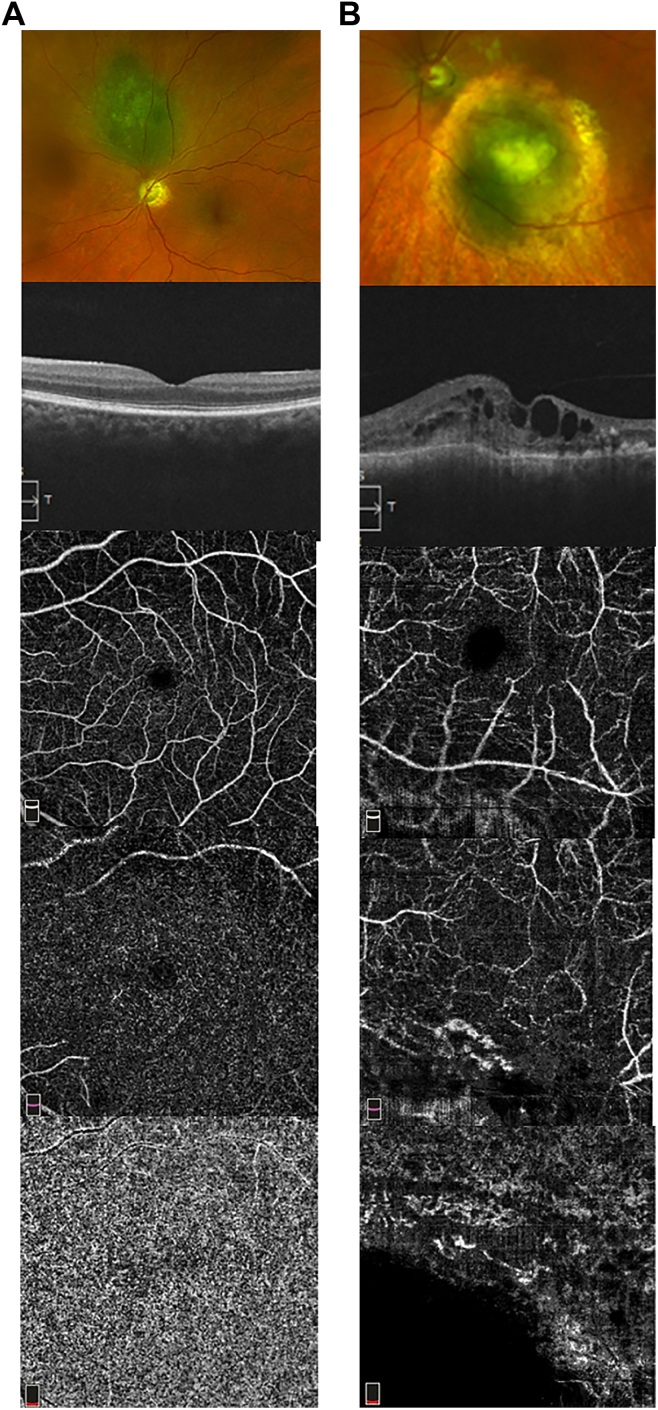
Purpose: To evaluate the macular and peripapillary retinal and choroidal flow changes in eyes with choroidal melanoma (CM) treated with proton beam radiation therapy (PBRT) using OCT angiography (OCTA).
Design: A prospective, cross-sectional, single-center study.
Participants: All patients seen at the study center between 2019 and 2024 who received PBRT for CM in 1 eye ≥1 year before enrollment with best-corrected visual acuity (BCVA) >20/200, unremarkable contralateral eye, and agreed to participate.
Methods: After a comprehensive eye examination, including BCVA, Optovue AngioVue was used to obtain the 4.5-mm optic disc and 6.0-mm macular OCT/OCT angiography (OCTA) images of both eyes. All vascular density (VD) measurements were obtained automatically using the OCTA software, except choriocapillaris VD, which was quantitated using ImageJ. The Wilcoxon signed-rank test was used to analyze differences in OCT/OCTA parameters between the treated and the contralateral eyes. Spearman's ρ was used to identify OCTA parameters associated with BCVA or radiation dose. A P value of <0.05 was considered statistically significant.
Main outcome measures: Foveal avascular zone (FAZ) area and perimeter, choriocapillaris and retinal (superficial and deep) capillary VD in the macula and radial peripapillary capillary (RPC) VD on OCTA; macular and retinal nerve fiber layer thickness on OCT, tumor location, laterality and size at baseline, BCVA of both eyes, PBRT dose, and duration of follow-up at enrollment.
Results: Among 24 participants, OCT/OCTA parameters were significantly different in the treated eyes when compared with the contralateral eyes, including increased FAZ area and perimeter, decreased peripapillary retinal nerve fiber layer thickness and RPC VD, and decreased macular choriocapillaris VD and parafoveal and perifoveal superficial retinal plexus VD (P < 0.05). Best-corrected visual acuity in the treated eyes correlated significantly with FAZ area and perimeter, parafoveal and perifoveal deep retinal plexus VD, and radiation dose to fovea but not radiation dose to the optic disc.
Conclusions: Although PBRT can affect both retinal and choroidal vascular flow in the macular and peripapillary region in eyes with CM, BCVA after PBRT seems to correlate best with the retinal vascular flow changes in the macula on OCTA and radiation dose to the fovea.
Financial disclosures: Proprietary or commercial disclosure may be found in the Footnotes and Disclosures at the end of this article.
Keywords: Choroid; Foveal avascular zone; Macula; Radiation; Uveal melanoma.
© 2024 by the American Academy of Ophthalmologyé.
Figures
Similar articles
-
Evaluating the Quantitative Foveal Avascular Zone and Retino-Choroidal Vessel Density Using Optical Coherence Tomography Angiography in a Healthy Indian Population.Cureus. 2022 Aug 4;14(8):e27669. doi: 10.7759/cureus.27669. eCollection 2022 Aug. Cureus. 2022. PMID: 36072178 Free PMC article.
-
Longitudinal Detection of Radiation-Induced Peripapillary and Macular Retinal Capillary Ischemia Using OCT Angiography.Ophthalmol Retina. 2020 Mar;4(3):320-326. doi: 10.1016/j.oret.2019.10.001. Epub 2019 Oct 11. Ophthalmol Retina. 2020. PMID: 31757690 Free PMC article.
-
[Correlation of capillary plexus with visual acuity in idiopathic macular epiretinal membrane eyes using optical coherence tomography angiography].Zhonghua Yan Ke Za Zhi. 2019 Oct 11;55(10):757-762. doi: 10.3760/cma.j.issn.0412-4081.2019.10.006. Zhonghua Yan Ke Za Zhi. 2019. PMID: 31607064 Chinese.
-
Microstructural and hemodynamic changes in the fundus after pars plana vitrectomy for different vitreoretinal diseases.Graefes Arch Clin Exp Ophthalmol. 2024 Jul;262(7):1977-1992. doi: 10.1007/s00417-023-06303-x. Epub 2023 Nov 20. Graefes Arch Clin Exp Ophthalmol. 2024. PMID: 37982887 Review.
-
Evaluating the Chorioretinal Microcirculation in Preeclampsia with OCT-Angiography: A Narrative Literature Review.J Clin Med. 2025 Jun 2;14(11):3913. doi: 10.3390/jcm14113913. J Clin Med. 2025. PMID: 40507675 Free PMC article. Review.
References
-
- Diener-West M., Reynolds S.M., Agugliaro D.J., et al. Screening for metastasis from choroidal melanoma: the collaborative ocular melanoma study group report 23. J Clin Oncol. 2004;22(12):2438–2444. - PubMed
-
- Shields J.A., Shields C.L. Management of posterior uveal melanoma: past, present, and future: the 2014 Charles L. Schepens lecture. Ophthalmology. 2015;122(2):414–428. - PubMed
-
- Matet A., Daruich A., Zografos L. Radiation maculopathy after proton beam therapy for uveal melanoma: optical coherence tomography angiography alterations influencing visual acuity. Invest Ophthalmol Vis Sci. 2017;58(10):3851–3861. - PubMed
LinkOut - more resources
Full Text Sources
