

Sex, senescence, senolytics, and cognition
- PMID: 40103928
- PMCID: PMC11913825
- DOI: 10.3389/fnagi.2025.1555872
Sex, senescence, senolytics, and cognition
Abstract
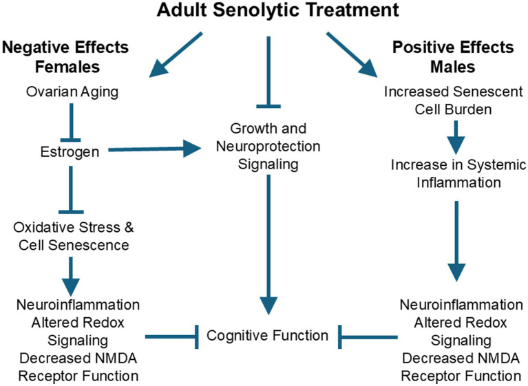
This review focuses on sexual dimorphism in cellular senescence and senolytic treatment in relation to brain health and age-related cognitive decline. The stressors of aging, DNA damage, inflammation, and oxidative stress induce cell senescence, a hallmark of aging. Senescent cells change their function and molecular profile and are primed to release pro-inflammatory cytokines. The functional changes include the activation of cell signals to prevent cell death. The release of pro-inflammatory cytokines from peripheral senescent cells during middle age induces senescence of neighbor cells and heightens the level of systemic inflammation, contributing to neuroinflammation. In response to neuroinflammation and oxidative stress, some neurons alter their physiology, decreasing neuronal excitability and synaptic transmission. Senescent neurophysiology is protective against cell death due to excitotoxicity, at the expense of a loss of normal cell function, contributing to age-related cognitive decline. The level of peripheral cell senescence and systemic inflammation may underlie sexual dimorphism in the prevalence, symptoms, and pathogenesis of age-related diseases, including neurodegenerative diseases. Sex differences have been observed for senescence of astrocytes, microglia, and peripheral cells, including those involved in innate and adaptive immune responses. Interventions that remove senescent cells, such as senolytic drugs, can reduce or ameliorate some of the aging-related loss of function. Similarities and differences in senolytic responses of males and females depend on the system examined, the treatment regimen, the level of senescent cell burden, and the age when treatment is initiated. Estrogen impacts several of these factors and influences the transcription of genes promoting growth, proliferation, and cell survival programs in a manner opposite that of senolytic drugs. In addition, estrogen has anti-aging effects that are independent of cell senescence, including rapidly modifying senescent neurophysiology. Thus, it is important to recognize that, in addition to sex differences in cell senescence, there are other sexually dimorphic mechanisms that contribute to the aging process. The results indicate that senolytics interact with fundamental biology, including sex hormones.
Keywords: aging; cellular senescence; senolytic treatment; sex differences; sex hormone.
Copyright © 2025 Foster and Kumar.
Conflict of interest statement
The authors declare that the research was conducted in the absence of any commercial or financial relationships that could be construed as a potential conflict of interest. The author(s) declared that they were an editorial board member of Frontiers, at the time of submission. This had no impact on the peer review process and the final decision.
Figures
References
Publication types
LinkOut - more resources
Full Text Sources