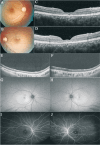
Angle closure glaucoma in a patient with X-linked retinoschisis: a case report
- PMID: 40103944
- PMCID: PMC11865655
- DOI: 10.18240/ijo.2025.03.24
Angle closure glaucoma in a patient with X-linked retinoschisis: a case report
Conflict of interest statement
Conflicts of Interest: Yao L, None; Lu Y, None; Yang KY, None; Lyu K, None; Wang ZY, None; Huang LZ, None; Wu HJ, None.
Figures


Similar articles
-
The treatment of refractory angle-closure glaucoma in a patient with X-linked juvenile retinoschisis.Ophthalmic Genet. 2018 Oct;39(5):625-627. doi: 10.1080/13816810.2018.1490961. Epub 2018 Aug 6. Ophthalmic Genet. 2018. PMID: 30081704
-
A paradigm shift in the treatment of refractory angle closure glaucoma in a patient with X-linked juvenile retinoschisis.Ophthalmic Genet. 2023 Dec;44(6):610-617. doi: 10.1080/13816810.2023.2188225. Epub 2023 Mar 16. Ophthalmic Genet. 2023. PMID: 36927170
-
Management of angle-closure glaucoma with X-linked retinoschisis: a case report.BMC Ophthalmol. 2023 Apr 17;23(1):159. doi: 10.1186/s12886-023-02903-7. BMC Ophthalmol. 2023. PMID: 37069516 Free PMC article.
-
[Papillomacular retinoschisis associated with glaucoma].Vestn Oftalmol. 2019;135(6):100-107. doi: 10.17116/oftalma2019135061100. Vestn Oftalmol. 2019. PMID: 32015314 Review. Russian.
-
Etiologies and clinical characteristics of young patients with angle-closure glaucoma: a 15-year single-center retrospective study.Graefes Arch Clin Exp Ophthalmol. 2021 Aug;259(8):2379-2387. doi: 10.1007/s00417-021-05172-6. Epub 2021 Apr 19. Graefes Arch Clin Exp Ophthalmol. 2021. PMID: 33876278 Free PMC article. Review.
References
-
- Hahn LC, van Schooneveld MJ, Wesseling NL, et al. X-linked retinoschisis: novel clinical observations and genetic spectrum in 340 patients. Ophthalmology. 2022;129(2):191–202. - PubMed
LinkOut - more resources
Full Text Sources
