

Refractive status and histological changes after posterior scleral reinforcement in guinea pig
- PMID: 40103945
- PMCID: PMC11865658
- DOI: 10.18240/ijo.2025.03.01
Refractive status and histological changes after posterior scleral reinforcement in guinea pig
Abstract
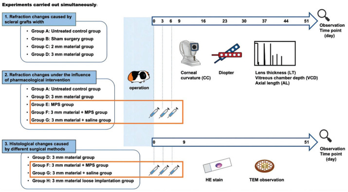
Aim: To investigate the refractive and the histological changes in guinea pig eyes after posterior scleral reinforcement with scleral allografts.
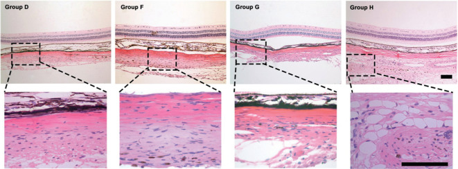
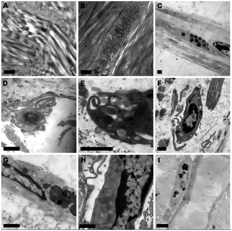
Methods: Four-week-old guinea pigs were implanted with scleral allografts, and the changes of refraction, corneal curvature and axis length were monitored for 51d. The effects of methylprednisolone (MPS) on refraction parameters were also evaluated. And the microstructure and ultra-microstructure of eyes were observed on the 9d and 51d after operation. Repeated-measures analysis of variance and one-way analysis of variance were used.
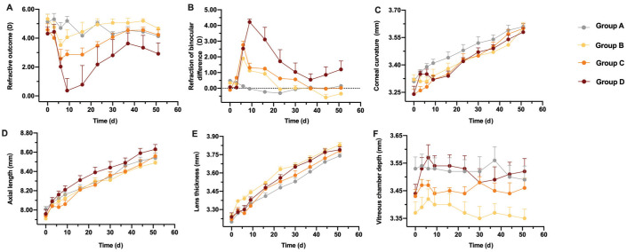
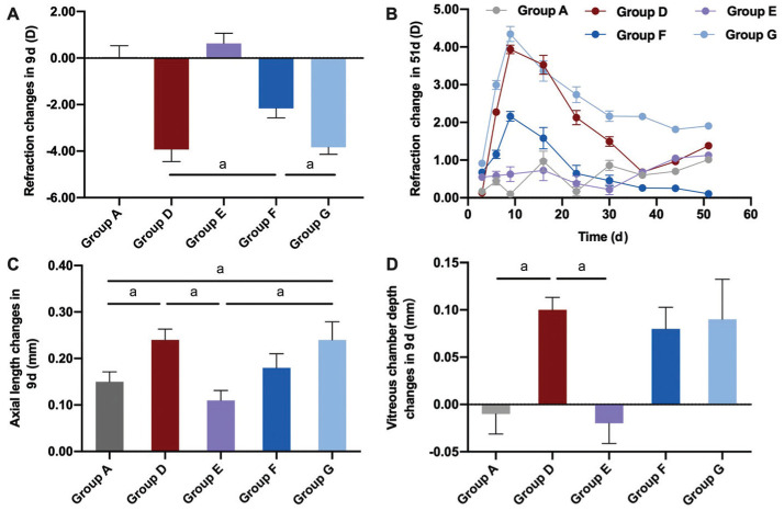
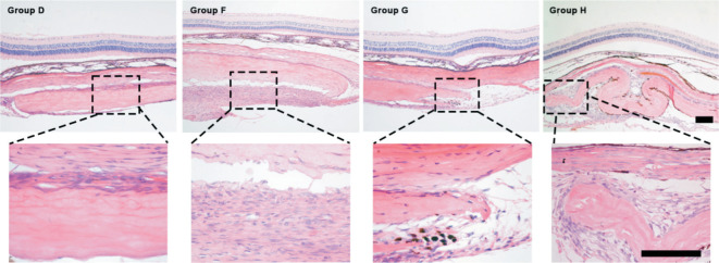
Results: The refraction outcome of the implanted eye decreased after operation, and the refraction change of the 3 mm scleral allografts group was significantly different with control group (P=0.005) and the sham surgical group (P=0.004). After the application of MPS solution, the reduction of refraction outcome was statistically suppressed (P=0.008). The inflammatory encapsulation appeared 9d after surgery. On 51d after operation, the loose implanted materials were absorbed, while the adherent implanted materials with MPS group were still tightly attached to the recipient's eyeball.
Conclusion: After implantation of scleral allografts, the refraction of guinea pig eyes fluctuated from a decrease to an increase. The outcome of the scleral allografts is affected by implantation methods and the inflammatory response. Stability of the material can be improved by MPS.
Keywords: guinea pig; inflammation; methylprednisolone; myopia; posterior scleral reinforcement.
International Journal of Ophthalmology Press.
Conflict of interest statement
Conflicts of Interest: Huang YY, None; Zhou LY, None; Chen GF, None; Peng D, None; Pan MZ, None; Zhou JB, None; Qu J, None.
Figures
Similar articles
-
Effects of scleral cross-linking using genipin on the process of form-deprivation myopia in the guinea pig: a randomized controlled experimental study.BMC Ophthalmol. 2015 Jul 29;15:89. doi: 10.1186/s12886-015-0086-z. BMC Ophthalmol. 2015. PMID: 26220299 Free PMC article.
-
Cytokine Expression and Biomechanical Characteristics after Posterior Scleral Reinforcement Using Demineralized Bone Matrix and Allogeneic Sclera.Altern Ther Health Med. 2024 Dec;30(12):486-494. Altern Ther Health Med. 2024. PMID: 38551429
-
Comparing the Differences in Slowing Myopia Progression by Riboflavin/Ultraviolet A Scleral Cross-linking before and after Lens-induced Myopia in Guinea Pigs.Curr Eye Res. 2022 Apr;47(4):531-539. doi: 10.1080/02713683.2021.2011324. Epub 2021 Dec 22. Curr Eye Res. 2022. PMID: 34935578
-
[Experimental study of glyceraldehyde cross-linking of posterior scleral on FDM in guinea pigs].Zhonghua Yan Ke Za Zhi. 2014 Jan;50(1):51-9. Zhonghua Yan Ke Za Zhi. 2014. PMID: 24709134 Chinese.
-
Treatment effect of posterior scleral reinforcement on controlling myopia progression: A systematic review and meta-analysis.PLoS One. 2020 May 26;15(5):e0233564. doi: 10.1371/journal.pone.0233564. eCollection 2020. PLoS One. 2020. PMID: 32453804 Free PMC article.
References
-
- Xue AQ, Bao FJ, Zheng LY, et al. Posterior scleral reinforcement on progressive high myopic young patients. Optom Vis Sci. 2014;91(4):412–418. - PubMed
-
- Ye J, Wu Y, Zhu SQ, et al. Evaluation of the efficacy of posterior scleral contraction in the treatment of macular hole with retinal detachment in high myopia. Retina. 2021;41(9):1874–1882. - PubMed
-
- Zheng LY, Pan AP, Zhu SQ, et al. Posterior scleral contraction to treat recurrent or persistent macular detachment after previous vitrectomy in highly myopic eyes. Retina. 2019;39(1):193–201. - PubMed
-
- Xue AQ, Zheng LY, Tan GL, et al. Genipin-crosslinked donor sclera for posterior scleral contraction/reinforcement to fight progressive myopia. Invest Ophthalmol Vis Sci. 2018;59(8):3564–3573. - PubMed
LinkOut - more resources
Full Text Sources