

Changes in retinal capillary density in female with type 2 diabetes and gestational diabetes-an analysis based on OCTA technology
- PMID: 40103950
- PMCID: PMC11865663
- DOI: 10.18240/ijo.2025.03.10
Changes in retinal capillary density in female with type 2 diabetes and gestational diabetes-an analysis based on OCTA technology
Abstract
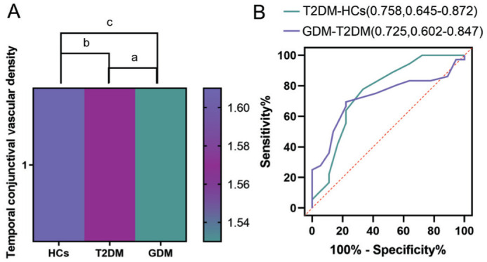
Aim: To evaluate alterations in conjunctival vascular density (CVD) and macular capillary density (MCD) in female patients with type 2 diabetes mellitus (T2DM) and gestational diabetes mellitus (GDM) using optical coherence tomography angiography (OCTA).
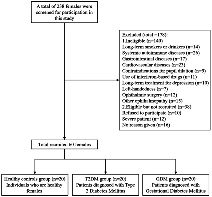
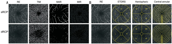
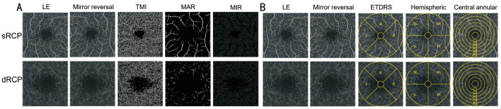
Methods: A total of 60 female participants were recruited, comprising 20 patients with T2DM, 20 patients with GDM, and 20 healthy age-matched controls (HCs). OCTA was used to assess superficial and deep retinal and conjunctival capillary plexuses. Subsequently, changes in MCD were analyzed using a circular segmentation method (C1-C6), a hemispheric quadrant segmentation method [superior right (SR), superior left (SL), inferior left (IL), and inferior right (IR)], and the early treatment diabetic retinopathy study (ETDRS) segmentation method (S, I, R, L).
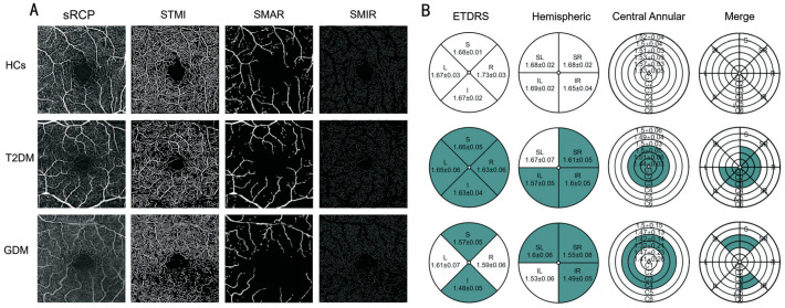
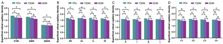
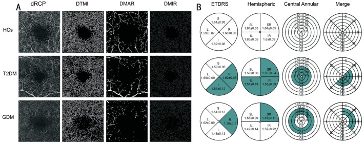
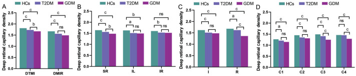
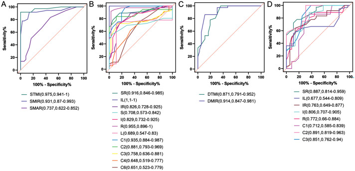
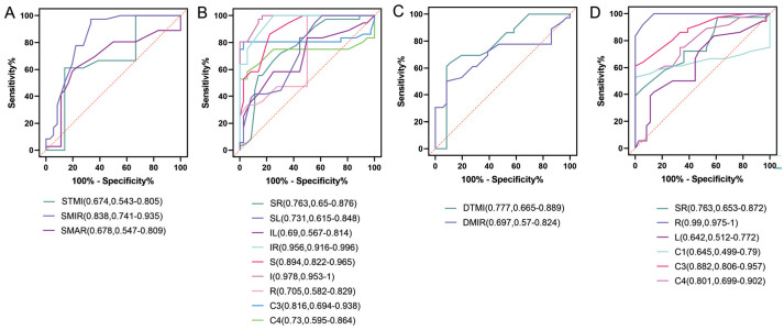
Results: OCTA unequivocally demonstrated that the variations in CVD among HCs, T2DM, and GDM groups were statistically significant (P<0.001). In the superficial retinal capillary plexus (sRCP), significant differences were observed in the densities of total microvascular (TMI), microvasculature (MIR), and macrovascular (MAR) between patients with T2DM and HCs (P<0.05). Furthermore, the GDM group exhibited a more substantial reduction in MIR density compared to the T2DM group (P<0.01). In the deep retinal capillary plexus (dRCP), significant differences in the densities of TMI and MIR were identified between the T2DM group and HCs (P<0.05), with a notable difference in TMI density also observed between the GDM and T2DM groups (P<0.01). In the receiver operating characteristic (ROC) curve analysis, the area under the ROC curve (AUC) for TMI in sRCP between the T2DM group and HCs was 0.975, with a 95% confidence interval (CI) of 0.941-1. The AUC for MIR was highest in dRCP, with an AUC value of 0.914 and a 95%CI ranging from 0.847 to 0.981. In comparing the GDM and T2DM groups, the AUC for I region was maximized in sRCP, achieving a value of 0.978 with a 95%CI of 0.953-1. Additionally, the AUC for R region was maximized in dRCP, reaching a value of 0.99 with a 95%CI of 0.975 to 1.
Conclusion: The sRCP and dRCP densities show higher diagnostic sensitivity for T2DM and GDM. OCTA holds potential as a significant instrument for the early diagnosis and differentiation of T2DM and GDM.
Keywords: deep retinal capillary plexus; diabetes; optical coherence tomography angiography; retinal region segmentation method; superficial retinal capillary plexus.
International Journal of Ophthalmology Press.
Conflict of interest statement
Conflicts of Interest: Zhang J, None; Yu Y, None; Zhou XM, None; Liao X, None; Hu JY, None; Ling Q, None; Zou J, None; Chen C, None; He LQ, None; Wei H, None; Chen X, None; Wang YX, None; Shao Y, None; Li RM, None.
Figures
Similar articles
-
OCTA evaluates changes in retinal microvasculature in renal hypertension patients.Sci Rep. 2024 Nov 21;14(1):28910. doi: 10.1038/s41598-024-68690-3. Sci Rep. 2024. PMID: 39572632 Free PMC article.
-
Microvascular alterations of the ocular surface and retina in connective tissue disease-related interstitial lung disease.Int J Ophthalmol. 2024 Oct 18;17(10):1869-1879. doi: 10.18240/ijo.2024.10.14. eCollection 2024. Int J Ophthalmol. 2024. PMID: 39430022 Free PMC article.
-
Retinal Microvasculature and Conjunctival Vessel Alterations in Patients With Systemic Lupus Erythematosus-An Optical Coherence Tomography Angiography Study.Front Med (Lausanne). 2021 Dec 2;8:724283. doi: 10.3389/fmed.2021.724283. eCollection 2021. Front Med (Lausanne). 2021. PMID: 34926488 Free PMC article.
-
Ocular microvascular alteration in patients with myocardial infarction-a new OCTA study.Sci Rep. 2024 Feb 24;14(1):4552. doi: 10.1038/s41598-023-50283-1. Sci Rep. 2024. PMID: 38402285 Free PMC article.
-
Evaluation of Posterior Ocular Blood Flow in Diabetic Retinopathy Patients Without Macular Edema Using Optical Coherence Tomography Angiography.Photodiagnosis Photodyn Ther. 2023 Dec;44:103777. doi: 10.1016/j.pdpdt.2023.103777. Epub 2023 Sep 3. Photodiagnosis Photodyn Ther. 2023. PMID: 37669724 Review.
References
-
- Harreiter J, Roden M. Diabetes mellitus—definition, classification, diagnosis, screening and prevention (update 2019) Wien Klin Wochenschr. 2019;131(Suppl 1):6–15. - PubMed
-
- Nanda M, Sharma R, Mubarik S, et al. Type-2 diabetes mellitus (T2DM): spatial-temporal patterns of incidence, mortality and attributable risk factors from 1990 to 2019 among 21 world regions. Endocrine. 2022;77(3):444–454. - PubMed
LinkOut - more resources
Full Text Sources
Research Materials