

Central alterations of brain networks in patients with optic neuritis: a resting state fMRI study
- PMID: 40103952
- PMCID: PMC11865650
- DOI: 10.18240/ijo.2025.03.14
Central alterations of brain networks in patients with optic neuritis: a resting state fMRI study
Abstract
Aim: To assess the alterations in the resting-state function connections between the two cerebral hemispheres in patients with optic neuritis (ON) and healthy controls (HCs).
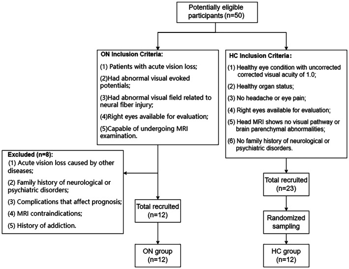
Methods: A total of 12 ON patients (six males and six females) and 12 HCs (six males and six females) who were highly matched for sex, age, and educational level were recruited. They underwent functional magnetic resonance imaging (fMRI), testing and brain activities were assessed using the degree centrality (DC) method. Correlation analysis between the mean DC values in specific brain areas and behavior performances was analyzed as well. Linear correlations between A anxiety scale (AS) and depression scale (DS) values and DC values in brain regions of patients with ON were also analyzed.
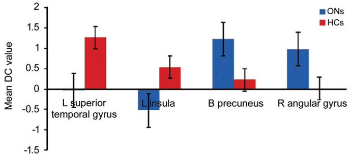
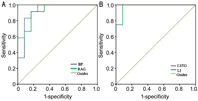
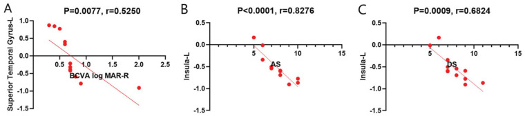
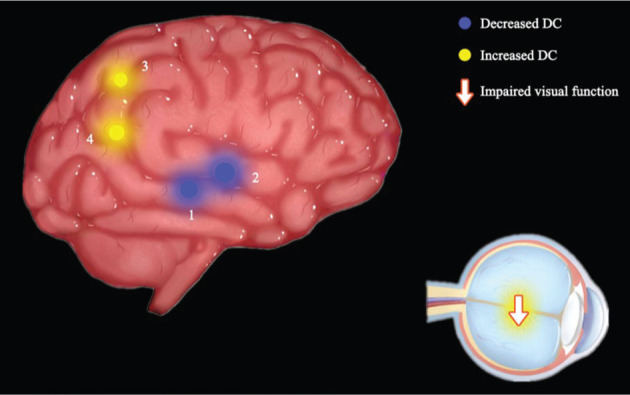
Results: The areas that showed a higher DC value in ON patients were the right angular gyrus and bilateral precuneus, while the left insula and left superior temporal gyrus (LSTG) were regions that presented a lower DC value in ON patients. A receiver operating characteristic (ROC) curve analysis confirmed the accuracy of the area under the curve (AUC) assessment. Linear analysis showed anxiety scale (AS) and depression scale (DS) values in the left insula were both negatively correlated with DC values, while best corrected visual acuity logMAR-R (BCVA logMAR-R) showed a negative correlation with DC in the LSTG.
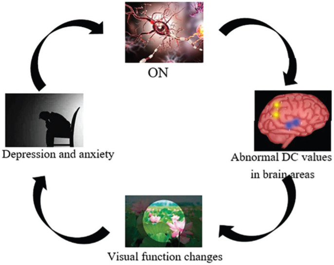
Conclusion: The study explores altered brain activities of specific regions in patients with ON. The results provide clues for revealing the underlying mechanism of ON development.
Keywords: functional magnetic resonance imaging; optic neuritis; resting state.
International Journal of Ophthalmology Press.
Conflict of interest statement
Conflicts of Interest: Huang L, None; Song D, None; Zhong L, None; Liao X, None; Zhou XM, None; Ge QM, None; Ling Q, None; Zeng YM, None; Wang XY, None; Hu JY, None; Chen C, None; He LQ, None; Zhou Q, None; Shao Y, None.
Figures

Similar articles
-
Spontaneous functional changes in specific cerebral regions in patients with hypertensive retinopathy: a resting-state functional magnetic resonance imaging study.Aging (Albany NY). 2021 May 10;13(9):13166-13178. doi: 10.18632/aging.202999. Epub 2021 May 10. Aging (Albany NY). 2021. PMID: 33972462 Free PMC article.
-
Altered Brain Network Centrality in Patients with Diabetic Optic Neuropathy: A Resting-State FMRI Study.Endocr Pract. 2020 Dec;26(12):1399-1405. doi: 10.4158/EP-2020-0045. Endocr Pract. 2020. PMID: 33471731
-
Alternations of interhemispheric functional connectivity in patients with optic neuritis using voxel-mirrored homotopic connectivity: A resting state fMRI study.Brain Imaging Behav. 2023 Feb;17(1):1-10. doi: 10.1007/s11682-022-00719-5. Epub 2022 Nov 28. Brain Imaging Behav. 2023. PMID: 36437427
-
Changes in Functional Connectivity of Specific Cerebral Regions in Patients with Toothache: A Resting-State Functional Magnetic Resonance Imaging Study.Dis Markers. 2020 Dec 28;2020:6683161. doi: 10.1155/2020/6683161. eCollection 2020. Dis Markers. 2020. PMID: 33456630 Free PMC article.
-
Alterations in degree centrality and functional connectivity in tension-type headache: a resting-state fMRI study.Brain Imaging Behav. 2024 Aug;18(4):819-829. doi: 10.1007/s11682-024-00875-w. Epub 2024 Mar 21. Brain Imaging Behav. 2024. PMID: 38512647 Review.
References
-
- Önder T, Karaçin C. Trastuzumab-induced optic neuritis: “blindness” side effect. J Oncol Pharm Pract. 2024:10781552241275538. - PubMed
-
- Lee JY, Han JN, Yang M, et al. Population-based incidence of pediatric and adult optic neuritis and the risk of multiple sclerosis. Ophthalmology. 2020;127(3):417–425. - PubMed
LinkOut - more resources
Full Text Sources
Research Materials
Miscellaneous