. 2025 Mar 18;15(11):8430-8442.
doi: 10.1039/d5ra00010f. eCollection 2025 Mar 17.

A calcium sulfate hemihydrate self-setting interface reinforced polycaprolactone porous composite scaffold

Affiliations

A calcium sulfate hemihydrate self-setting interface reinforced polycaprolactone porous composite scaffold

Changfeng Li et al. RSC Adv. .

Abstract

The mechanical insufficiency and slow degradation of polycaprolactone (PCL) implants have attracted widespread attention among researchers. Herein, a PCL scaffold with self-setting properties containing calcium sulfate hemihydrate (CSH) was prepared using a triply periodic minimal surfaces (TPMS) design and selective laser sintering (SLS) technology. The results showed that the strength of the scaffold containing 10 wt% CSH was increased by 45.5% compared to the PCL one. More importantly, its strength can be further increased to 1.7 times that of the PCL scaffold after self-setting in water. Mechanism analysis suggests that mechanical strengthening can be attributed to the pinning effect through the newly grown columnar crystals embedded with PCL molecular chains. In addition, the degradation rate of the composite scaffold was approximately 6.8 times higher than that of the PCL one. The study believes that the increase in degradation rate is due to a dual effect, specifically the increase in permeability and the catalytic degradation of PCL in the acidic environment. Encouragingly, the composite scaffold showed a good ability to induce hydroxyapatite formation. Therefore, the self-setting mechanically enhanced composite scaffold is expected to have potential application prospects in bone defect repair.


Conflict of interest statement

The authors declare that they have no known competing financial interests or personal relationships that could have appeared to influence the work reported in this paper.

Figures

Fig. 1
Fig. 1. Flow chart of scaffolds and powders preparation. (a) Preparation flow chart; (b) morphology of the prepared scaffold.
Fig. 2
Fig. 2. Characterization of powders and scaffolds. (a) SEM image of PCL; (b) SEM image of CSH; (c) PCL particle size distribution; (d) CSH particle size distribution; (e) XRD pattern of CSH; (f) XRD patterns of the scaffolds. (g) FTIR spectra of CSH and scaffolds. (h) XRD patterns of the scaffolds after soaking. (i) Porosity of the scaffold before and after soaking.
Fig. 3
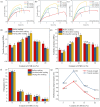
Fig. 3. Tensile properties of different scaffolds. Stress–strain curves of (a) before soaking and (b) not dried after soaking and (c) drying after soaking; (d) strength; (e) modulus; (f) elongation; (g) performance improvement.
Fig. 4
Fig. 4. Compression properties of the different scaffolds at 40% strain. Stress–strain curves of (a) before soaking and (b) not dried after soaking and (c) drying after soaking; (d) strength; (e) modulus.
Fig. 5
Fig. 5. Microscopic surface morphology of different scaffolds.
Fig. 6
Fig. 6. Tensile fracture morphology of different scaffolds.
Fig. 7
Fig. 7. Self-setting enhancement mechanical mechanism.
Fig. 8
Fig. 8. Changes in degradation properties of different scaffolds. (a) Water contact angle; (b) water absorption; (c) weight loss of the scaffolds; (d) pH of the solution.
Fig. 9
Fig. 9. Microscopic morphologies of different scaffolds after soaking in PBS.
Fig. 10
Fig. 10. Changes in soaking time and mechanical properties of different scaffolds. (a) Tensile stress–strain curve after soaking for 1 week and (b) soaking for 2 weeks; (c) tensile strength change; (d) compressive stress–strain curve after soaking for 1 week and (e) soaking for 2 weeks; (f) compressive strength change.
Fig. 11
Fig. 11. Surface micromorphology and XRD patterns of the scaffolds after soaking in SBF for 7 days. (a) PCL-5% CSH, (b) PCL-10% CSH, (c) PCL-15% CSH, (d) PCL-20% CSH, and (e) PCL represent the microscopic morphologies of the scaffolds. (f) XRD patterns of the scaffolds.

Similar articles

References

    1. Chen Y. Quan S. Huang S. Liu W. Chen Z. Liu J. Li C. Yang H. Ceram. Int. 2024;50:48891–48908.
    1. Sheng X. Che Z. Qiao H. Qiu C. Wu J. Li C. Tan C. Li J. Wang G. Liu W. Gao H. Li X. Int. J. Biol. Macromol. 2024;277:133806. - PubMed
    1. Li L. Shi J. Ma K. Jin J. Wang P. Liang H. Cao Y. Wang X. Jiang Q. J. Adv. Res. 2021;30:75–84. - PMC - PubMed
    1. Kumar Parupelli S. Saudi S. Bhattarai N. Desai S. Int. J. Bioprint. 2023;9:0196.
    1. Zhao X. Li N. Zhang Z. Hong J. Zhang X. Hao Y. Wang J. Xie Q. Zhang Y. Li H. Liu M. Zhang P. Ren X. Wang X. J. Nanobiotechnol. 2024;22:500. - PMC - PubMed

LinkOut - more resources