. 2025 Feb 28:48:510-528.
doi: 10.1016/j.bioactmat.2025.02.034. eCollection 2025 Jun.

Multifunctional magneto-electric and exosome-loaded hydrogel enhances neuronal differentiation and immunoregulation through remote non-invasive electrical stimulation for neurological recovery after spinal cord injury

Affiliations

Multifunctional magneto-electric and exosome-loaded hydrogel enhances neuronal differentiation and immunoregulation through remote non-invasive electrical stimulation for neurological recovery after spinal cord injury

Wubo Liu et al. Bioact Mater. .

Abstract

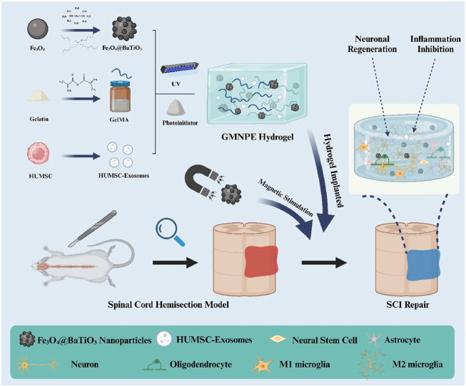
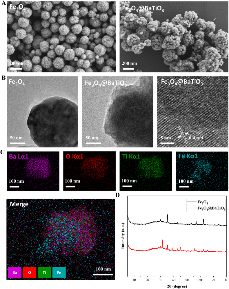
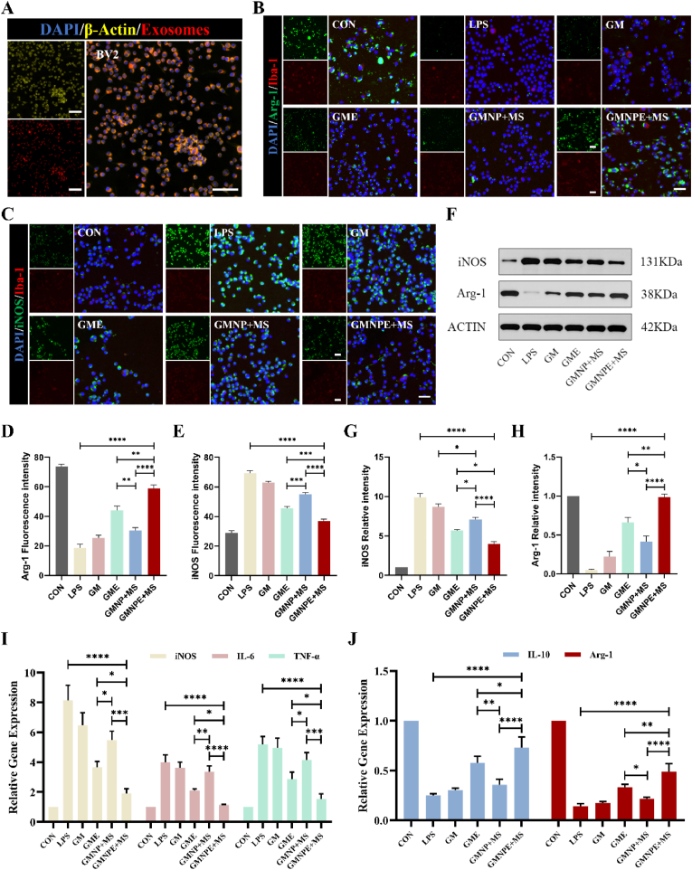
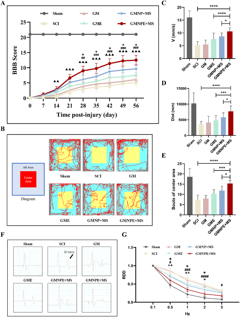
Intervention in the differentiation of neural stem cells (NSCs) is emerging as a highly promising approach for the treatment of spinal cord injury (SCI). However, NSCs at the injury site often suffer from low survival and uncontrolled differentiation. Whereas electrical stimulation has proven effective in regulating the fate of NSCs and promoting tissue repair, however, conventional electrical stimulation therapy has failed to be widely applied due to challenges such as invasiveness and technical complexity. To overcome these limitations, we developed a biomimetic magneto-electric hydrogel incorporating Fe3O4@BaTiO3 core-shell nanoparticles and human umbilical mesenchymal stem cell exosomes (HUMSC-Exos) around the concept of constructing remote noninvasive electrical stimulation for the synergistic treatment of SCI. The Fe3O4@BaTiO3 is activated by the peripheral magnetic field to generate electrical stimulation, which, in conjunction with the synergistic effects of HUMSC-Exos, significantly alleviates the early inflammatory response associated with SCI and enhances the regeneration of newborn neurons and axons, thereby creating favorable conditions for functional recovery post-SCI. Our findings indicate that applying this magneto-exosome hydrogel in a rat model of SCI leads to substantial functional recovery. This innovative combination represents a promising therapeutic strategy for SCI repair.

Keywords: HUMSC-Exos; Magneto-electric nanoparticles; Neuronal differentiation; Spinal cord injury repair; Tissue engineering.


Conflict of interest statement

The authors declare no conflict of interest.

Figures

Image 1
Graphical abstract
Scheme 1
Scheme 1
Schematic illustration of the synthesis steps of GMNPE hydrogel and the promotion of neuronal regeneration after SCI as well as inflammation inhibition in the presence of a remote magnetic field.
Fig. 1
Fig. 1
Characterization of Fe3O4@BaTiO3 nanoparticles. (A) SEM images of Fe3O4 and Fe3O4@BaTiO3 nanoparticles. (B) TEM images of Fe3O4 and Fe3O4@BaTiO3 nanoparticles, and high-resolution TEM image of Fe3O4@BaTiO3 nanoparticles. (C) EDX mapping of Fe3O4@BaTiO3 nanoparticles. (D) XRD result of Fe3O4 and Fe3O4@BaTiO3 nanoparticles.
Fig. 2
Fig. 2
Characterization of the HUMSC-Exos and GMNPE hydrogels. (A) The analysis results of Flow NanoAnalyzer showing exosomes with a particle size of 81.1 nm (gating range 47.1–190.0 nm). (B) TEM image revealed the characteristic cup shape (white arrow) of the exosomes. (C) WB results showing that exosomes positively expressed CD9, CD81, and Alix, along with the absence of the negative marker Calnexin. (D) different states of hydrogels: GM hydrogel before gel formation (left), GMNPE hydrogel before gel formation (middle), GMNPE hydrogel after gel formation (right). (E) SEM images of GM hydrogel and GMNPE hydrogel at low and high magnification. Scale bar: 100 μm. Scale bar of magnified image: 1 μm. (F) EDX mapping of GMNPE hydrogel. (G) FTIR spectra of the Fe3O4@BaTiO3, GM hydrogel, GM + LAP hydrogel, and GMNPE hydrogel. (H) The output current of GMNPE hydrogel under magnetic field stimulation (5 mT, 10 mT and 15 mT). (I) The mechanistic properties of GM and GMNPE hydrogels. (J) Swelling properties of GM and GMNPE hydrogels (n = 4).
Fig. 3
Fig. 3
Anti-inflammatory effects of GMNPE in vitro. (A) BV2 cells cultured on GMNPE hydrogels can normally phagocytose PKH26-stained HUMSC-Exos released from hydrogels. Scale bars: 100 μm. (B–C) Immunofluorescence images of BV2 cells cultured in each group. Cells were stained red with ionized calcium-binding adaptor molecule 1, and Arg-1/iNOS was stained green. Scale bar: 50 μm. (D–E) Quantification of fluorescence intensity of Arg-1 and iNOS level in each group (n = 5). (F) Western blot images showed the changes in protein expression of iNOS and Arg-1. (G–H) Quantitative analysis of iNOS/Actin and Arg-1/Actin ratio (n = 3). (I) RT-qPCR results of the pro-inflammatory cytokines iNOS, IL-6 and TNF-α (n = 3). (J) RT-qPCR results of the anti-inflammatory cytokines Arg-1 and IL-10 (n = 3). Significance: ns-not significant, ∗p < 0.05, ∗∗p < 0.01, ∗∗∗p < 0.001, ∗∗∗∗p < 0.0001.
Fig. 4
Fig. 4
NSCs differentiation and axon regeneration on hydrogels. (A) An illustration of GMNPE hydrogel promoting NSCs differentiation to neurons and oligodendrocytes and axon regeneration. (B) Immunofluorescence staining of NSCs cultured on different groups. Red IF represents the neuron marker Tuj-1, astrocyte marker GFAP, oligodendrocyte marker MBP, or neurofilament marker NF respectively. Scale bar: Tuj-1, GFAP, MBP, 50 μm. NF, 150 μm. (C–E) Statistical analysis of the percentage of Tuj-1 (+), GFAP (+) and MBP (+) cells to total cell number (n = 5). (F–G) Statistical analysis of the NF (+) area ratio and the length of NF (+) axon (n = 5). (H) Western blot images showed the changes in protein expression of Tuj-1, GFAP, MBP and NF. (I–L) Quantitative analysis of Tuj-1/ACTIN, GFAP/ACTIN, MBP/ACTIN and NF/ACTIN ratio in Western blot images (n = 3). (M) RT-qPCR analysis of the gene expression of Tuj1, GFAP, MBP and NF (n = 3). Significance: ∗p < 0.05, ∗∗p < 0.01, ∗∗∗p < 0.001, ∗∗∗∗p < 0.0001.
Fig. 5
Fig. 5
The mechanism of GMNPE + MS regulating the differentiation of NSCs and axon regeneration. (A) Heatmap illustrating differential genes in the GMNPE + MS group compared to the CON group (log2 Fold change ≥0.5 and q < 0.05). (B) Presentation of the top 15 entries in the bubble diagram of GO enrichment (n = 3). (C) The bubble diagram of KEGG enrichment (n = 3). (D) Western blot images showed the expression of the PI3K-AKT pathway protein between the CON group and the GMNPE + MS group.
Fig. 6
Fig. 6
Functional recovery of rats in different groups. (A) The BBB score of rats in each group (n = 8). (B) An illustration of the open field test. (C–E) Quantitative analysis of distance, speed and bouts of center area for each group in the open field test (n = 8). (F) Electrophysiological detection waveforms of each group. The black arrow represents the H-wave. (G) Results of the analysis of electrophysiological RDD data at 8 weeks after SCI in each group (n = 5). Symbol (∗) represented the comparison of the GMNPE + MS group with the GMNP + MS group. Symbol (#) represented the comparison of the GMNPE + MS group with the GME group. Symbol (▲) represented the comparison of the GMNPE + MS group with the SCI group. Symbol (+) represented the comparison of the GMNPE + MS group with the Sham group. Significance: ns-not significant, ∗p < 0.05, ∗∗p < 0.01, ∗∗∗p < 0.001, ∗∗∗∗p < 0.0001.
Fig. 7
Fig. 7
GMNPE hydrogel inhibited the inflammatory microenvironment in the early stage after SCI. (A) Representative images of immunofluorescence staining of CD68 in each group at day 7 after SCI. Scale bar: 100 μm. (B) Statistical analysis of CD68 (+) cell percentage to total cell number. (n = 3). (C) Western blot image showed the changes in protein expression of iNOS and Arg-1. (D–E) Quantitative analysis of iNOS/ACTIN and Arg-1/ACTIN ratio in western blot images (n = 3). Significance: ns-not significant, ∗p < 0.05, ∗∗p < 0.01, ∗∗∗p < 0.001, ∗∗∗∗p < 0.0001.
Fig. 8
Fig. 8
GMNPE + MS induced differentiation of endogenous NSCs into functional neurons and promoted neuronal regeneration. (A) Representative images of immunofluorescence staining of Tuj-1 (green) and GFAP (red) in each group at week 8 after SCI. Scale bar: 200 μm. Scale bar of magnified image: 50 μm. (B–C) The ratio of Tuj-1 (+) area and GFAP (+) area were quantified (n = 3). (D) Representative images of immunofluorescence staining of MBP (green) and NF (red) in each group at week 8 after SCI. Scale bars: 50 μm. (E–F) The ratio of MBP (+) area and NF (+) area were quantified (n = 3). (G) Western blot image showed the changes in protein expression of Tuj-1 and GFAP. (H–I) Quantitative analysis of Tuj-1/ACTIN and GFAP/ACTIN ratio in western blot images (n = 3). Significance: ns-not significant, ∗p < 0.05, ∗∗p < 0.01, ∗∗∗p < 0.001, ∗∗∗∗p < 0.0001.

Similar articles

Cited by

References

    1. Karsy M., Hawryluk G. Modern medical management of spinal cord injury. Curr. Neurol. Neurosci. Rep. 2019;19:65. - PubMed
    1. Quadri S.A., Farooqui M., Ikram A., Zafar A., Khan M.A., Suriya S.S., Claus C.F., Fiani B., Rahman M., Ramachandran A., Armstrong I.I.T., Taqi M.A., Mortazavi M.M. Recent update on basic mechanisms of spinal cord injury. Neurosurg. Rev. 2020;43:425–441. - PubMed
    1. Zhang C., Talifu Z., Xu X., Liu W., Ke H., Pan Y., Li Y., Bai F., Jing Y., Li Z., Li Z., Yang D., Gao F., Du L., Li J., Yu Y. MicroRNAs in spinal cord injury: a narrative review. Front. Mol. Neurosci. 2023;16 - PMC - PubMed
    1. Hutson T.H., Di Giovanni S. The translational landscape in spinal cord injury: focus on neuroplasticity and regeneration. Nat. Rev. Neurol. 2019;15:732–745. - PubMed
    1. Li X., Peng Z., Long L., Tuo Y., Wang L., Zhao X., Le W., Wan Y. Wnt4-modified NSC transplantation promotes functional recovery after spinal cord injury. FASEB J. 2020;34:82–94. - PubMed

LinkOut - more resources