. 2025 Feb 13;28(3):112021.
doi: 10.1016/j.isci.2025.112021. eCollection 2025 Mar 21.

Superior colliculus peri-saccadic field potentials are dominated by a visual sensory preference for the upper visual field

Affiliations

Superior colliculus peri-saccadic field potentials are dominated by a visual sensory preference for the upper visual field

Ziad M Hafed. iScience. .

Abstract

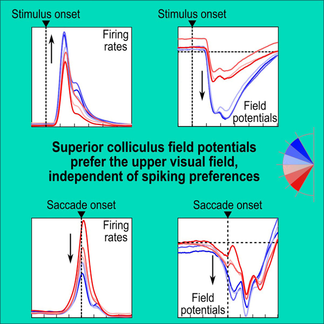
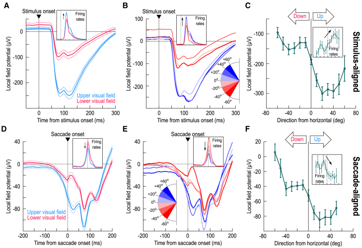
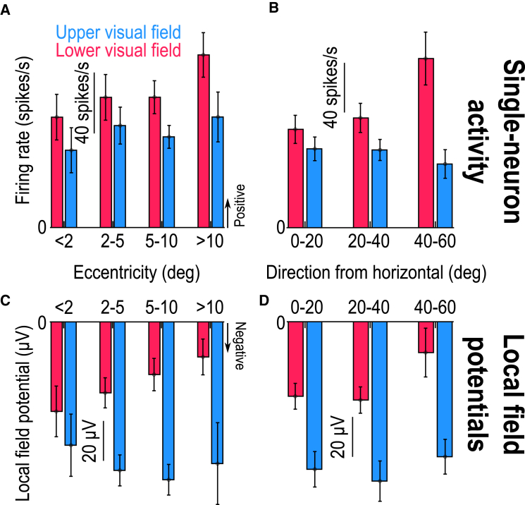
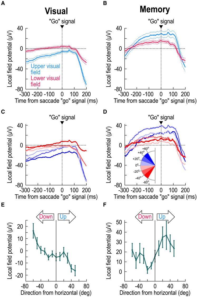
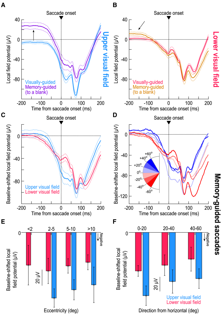
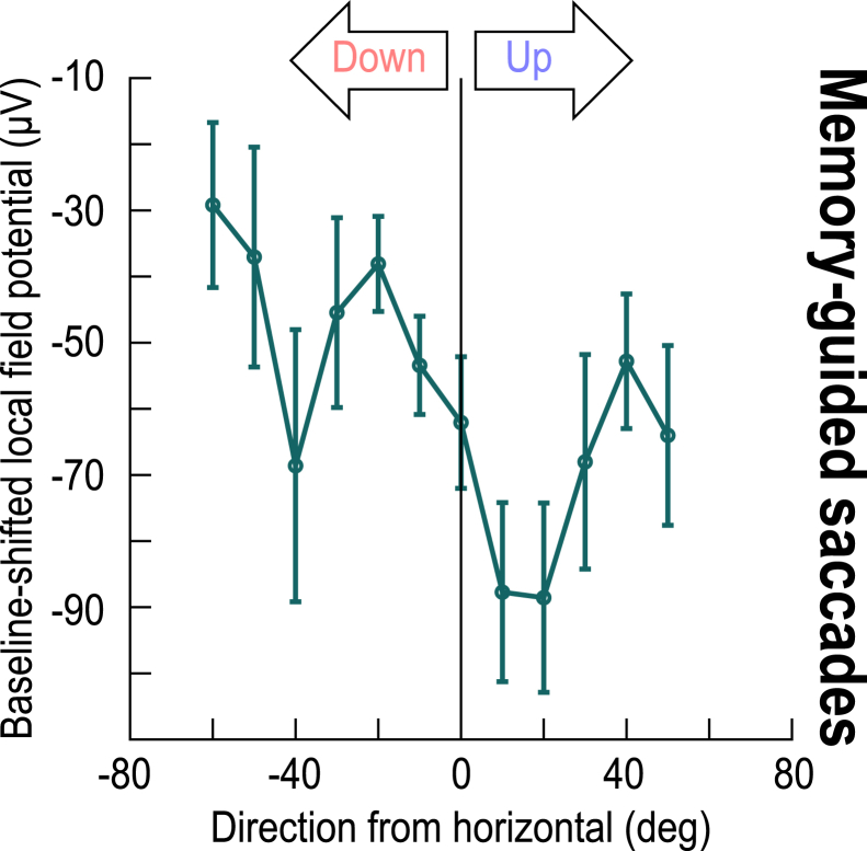
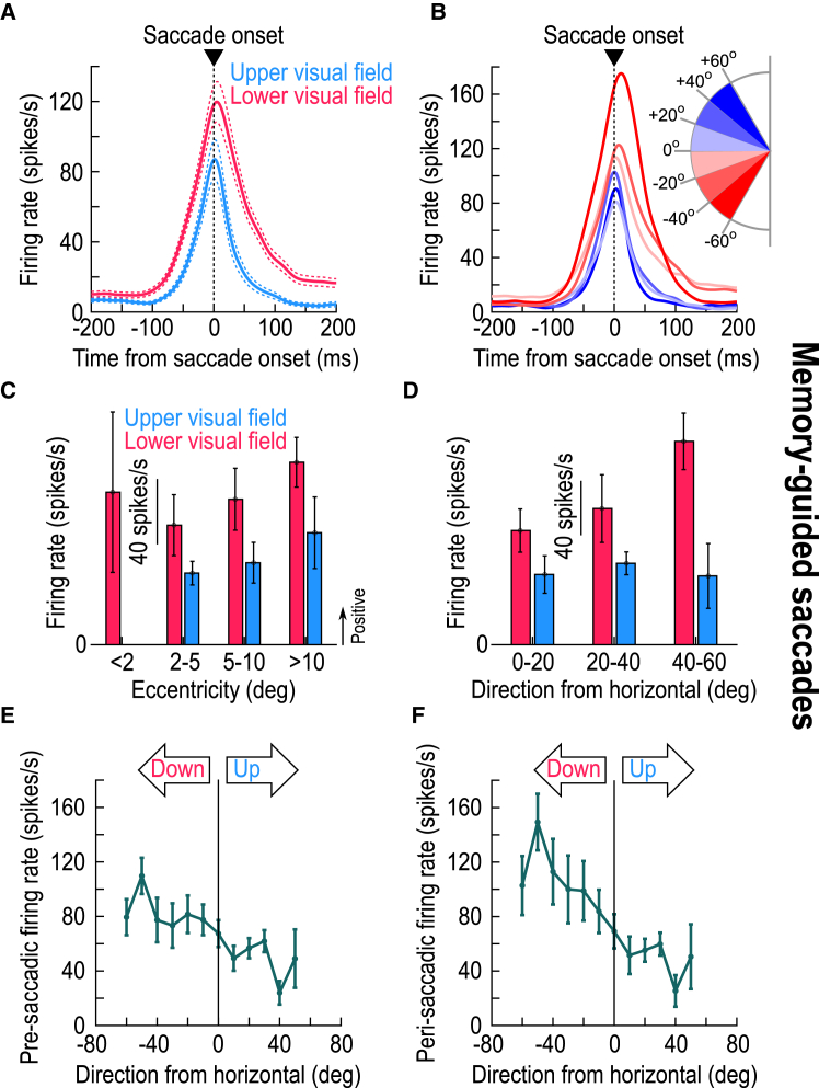
The primate superior colliculus (SC) plays important sensory, cognitive, and motor processing roles. Among its properties, the SC has clear visual field asymmetries: visual responses are stronger in the upper visual field representation, whereas saccade-related motor bursts are weaker. Here, I asked whether peri-saccadic SC network activity can still reflect the SC's visual sensitivity asymmetry, thus supporting recent evidence of sensory-related signals embedded within the SC's motor bursts. I analyzed collicular peri-saccadic local field potential (LFP) modulations and found them to be much stronger in the upper visual field, despite the weaker motor bursts. This effect persisted even with saccades toward a blank, suggesting an importance of visual field location. I also found that engaging working memory during saccade preparation differentially modulated the SC's LFP's, again with a dichotomous upper/lower visual field asymmetry. I conclude that the SC network possesses a clear sensory signal at the time of saccade generation.

Keywords: Cognitive neuroscience; Neuroscience; Sensory neuroscience.


Conflict of interest statement

The author declares no competing interests.

Figures

None
Graphical abstract
Figure 1
Figure 1
Peri-saccadic local field potential (LFP) deflections in the superior colliculus (SC) reflect their visual counterparts (A) Stimulus-evoked LFP deflections after the brief appearance of a small white spot at the preferred response field (RF) location. Blue shows the average for all electrode tracks in the SC’s upper visual field representation; red shows results from the lower visual field representation. The inset shows the corresponding single-neuron firing rates. LFP negativity deflections were much stronger in the upper visual field representation. (B and C) The same data but now separated as a function of direction from the horizontal axis represented by the SC sites. There was a step-like discontinuity across the horizon. (D) Similar to A but now for data aligned to saccade onset toward a visible spot. Even though the motor bursts were weaker in the upper visual field, (inset), the LFP negativity deflections were still much stronger for the upper visual field, consistent with the stimulus-evoked effects in A. (E and F) The same discontinuity across the horizontal meridian seen in B and C was also evident peri-saccadically (0–50 ms from saccade onset in F; Methods). Note how the insets in B and E as well as those in C and F show directly opposing dependencies in the spiking activity between the visual and motor epochs; in contrast, the LFP’s show the same dependencies. Error bars in all panels denote SEM. A–C were reproduced (using updated color schemes) with permission from our earlier study for easier direct visualization next to D–F. All insets were also reproduced from the same study.
Figure 2
Figure 2
Consistency of the results of Figure 1 across tested eccentricities and directions (A and B) For all single neurons from the database, I binned them according to the eccentricity (A) or direction from horizontal (B) of their RF hotspots (similar binning to what we had done with visually-evoked LFP measurements in our earlier study14). This confirmed the consistently weaker motor bursts in the upper visual field. In each bin, the measurements were made in the interval 0–50 ms from saccade onset. (C and D) For peri-saccadic LFP’s, there was always a stronger negativity for the upper visual field, like in the stimulus-evoked deflections (Figure 1) and opposite to the spiking asymmetry of A and B. Error bars denote SEM. Figures S1 and S2 document similar results when measuring pre-saccadic activity.
Figure 3
Figure 3
Both visual and working memory LFP representations depend on visual field location, but in diametrically opposite ways (A) Same as Figure 1D but aligned to the end of the delay period that existed between stimulus onset and the “go” signal for the saccade (in the delayed, visually-guided saccade task; Methods). Here, I aligned the data to the time of the go signal, when the fixation spot was removed and the eccentric spot (saccade target) was still visible. Before the end of the delay period, there was a sustained effect in which there was stronger LFP negativity for upper visual field SC sites. This is consistent with the results of Figures 1A–1C and suggests that continuous visual stimulation has persistent effects on the SC’s LFP’s. The strong negative deflections ∼150–200 ms after the go signal reflect the peri-saccadic LFP modulations of Figures 1D–1F. (B) In the absence of a visible saccade target, LFP’s turned positive after the initial negativity transient associated with the earlier stimulus onsets; thus, the SC’s LFP’s were clearly positive by the end of the delay period. Interestingly, there was still a dependence on the visual field location: SC sites representing the upper visual field had a larger LFP positivity during working memory than SC sites representing the lower visual field. Figure 4 shows that, despite this difference in LFP signals at the time of the go signal for memory-guided saccades, later peri-saccadic negativity amplitudes still behaved like those in the visually-guided saccade task. (C and D) Same as A, B, but now separating electrode tracks as a function of the direction from horizontal represented by the targeted SC sites. For the visual condition (C), the same sensory dependence of Figures 1A–1C was observed, again suggesting a sustained sensory influence on SC LFP’s. In the case of memory (D), there was clearly a visual field effect, but this time of the opposite sign: upper visual field SC sites had stronger LFP positivity, rather than negativity, at the end of the delay period. (E and F) Same as Figures 1C and 1F, but this time when measuring LFP values at the end of the delay period in both tasks (from −50 ms to +25 ms relative to the time of the saccade go signal; Methods). In both cases, there was an upper/lower visual field effect, but having different signs. Error bars denote SEM. Also see Figure S3.
Figure 4
Figure 4
Stronger upper visual field peri-saccadic LFP modulations even in the absence of a visible saccade target (A) Peri-saccadic LFP’s for upper visual field electrode tracks with and without a visible target (the visible target curve is the same as that in Figure 1D). Long before the saccade (upward arrow), the memory condition elevated LFP voltages (Figure 3). However, the peri-saccadic deflection magnitude was similar in the two curves. (B) Same as A but for the lower visual field. Here, the memory condition had a much weaker effect long before saccade onset (oblique arrow) (Figure 3). Importantly, the peri-saccadic deflection strength was still similar in both conditions. (C) Applying a baseline shift in the memory condition (Methods) revealed a similar peri-saccadic asymmetry between the upper and lower visual fields in the absence of a visible saccade target; there was stronger negativity in the upper visual field. (D) Same as Figure 1E but for the memory condition (after baseline shift). Upper visual field tracks had consistently stronger peri-saccadic LFP negativity. For example, compare the sites directly straddling the horizontal meridian (least saturated blue and least saturated red), especially in the peri-saccadic interval. (E and F) Same as Figures 2C and 2D, but for the memory condition, again showing stronger negativity in the upper visual field (the missing bar for <2 deg eccentricity means no data). Error bars in all panels indicate SEM.
Figure 5
Figure 5
Direction-dependence of peri-saccadic LFP modulations in the absence of a visible saccade target This figure bins all electrode tracks from Figure 4 as a function of the direction from horizontal represented by the recorded SC neurons. For baseline-shifted peri-saccadic LFP measurements (during the interval of 0–50 ms from memory-guided saccade onset), there was a stronger LFP negativity in the upper visual field representation of the SC than in the lower visual field representation. This was confirmed statistically when I grouped all upper visual field electrode tracks into one group and all lower visual field electrode tracks into another (p = 0.003; t = −3.0302; n = 49 and 81 for upper and lower visual field electrode tracks, respectively; t test). Similarly, a one-way ANOVA across the shown angular direction bins revealed a significant effect of direction from the horizontal meridian (p = 0.0123; F(11,222) = 2.27; n = 223). Error bars denote SEM.
Figure 6
Figure 6
Weaker motor bursts in the upper visual field in the absence of a visible saccade target (A and B) Like the insets of Figures 1D and 1E but for the memory-guided saccade task. Note how the firing rates long before saccade onset did not depend on visual field location, even though the LFP effects did (Figures 3, 4A, and 4B). This suggests another dissociation between SC LFP’s and spiking activity, beyond the dissociation I highlighted previously for peri-saccadic intervals (e.g., Figures 1, 2, S1, and S2). (C and D) Like Figures 2A and 2B but for memory-guided saccades. Motor bursts in the upper visual field were weaker than in the lower visual field, which is opposite the peri-saccadic LFP asymmetries (Figures 4 and 5). (E) Similar to Figures 1C, 1F, 3E, 3F, 5, and S1, except that I now measured firing rates in the final 50 ms before saccade onset. Pre-saccadic firing rates were weaker in the upper than in the lower SC visual field representation, and this happened even with saccades toward a blank (memory-guided saccades). I confirmed this with a t test comparing all upper visual field neurons in the database in one group to all lower visual field neurons in the other (p = 0.0039; t = −2.9369; n = 49 and 81 for upper and lower visual field electrode tracks, respectively). I also performed a one-way ANOVA across all shown angular direction bins, again with a significant effect (p = 0.0329; F(11,211) = 1.97; n = 223). (F) Same as (E) but now for the measurement interval 0–50 ms from saccade onset. Again, peri-saccadic firing rates were weaker in the upper visual field for memory-guided saccades (p = 0.0037; F(11,211) = 2.62; n = 223; one-way ANOVA across all shown angular direction bins). Note that the overall firing rates were generally higher than in (E) (same y axis ranges in the two panels), which is expected because motor bursts are known to reach peak firing rates some time peri-saccadically rather than pre-saccadically., Error bars denote SEM.

Similar articles

Cited by

References

    1. Robinson D.A. Eye movements evoked by collicular stimulation in the alert monkey. Vis. Res. 1972;12:1795–1808. - PubMed
    1. Gandhi N.J., Katnani H.A. Motor functions of the superior colliculus. Annu. Rev. Neurosci. 2011;34:205–231. doi: 10.1146/annurev-neuro-061010-113728. - DOI - PMC - PubMed
    1. Munoz D.P., Wurtz R.H. Saccade-related activity in monkey superior colliculus. I. Characteristics of burst and buildup cells. J. Neurophysiol. 1995;73:2313–2333. - PubMed
    1. Munoz D.P., Wurtz R.H. Saccade-related activity in monkey superior colliculus. II. Spread of activity during saccades. J. Neurophysiol. 1995;73:2334–2348. - PubMed
    1. Lee C., Rohrer W.H., Sparks D.L. Population coding of saccadic eye movements by neurons in the superior colliculus. Nature. 1988;332:357–360. doi: 10.1038/332357a0. - DOI - PubMed

LinkOut - more resources