. 2025 Jan 23;28(3):111880.
doi: 10.1016/j.isci.2025.111880. eCollection 2025 Mar 21.

ROS-responsive nanoparticles with selenomethionine for ferroptosis modulation in abdominal aortic aneurysm

Affiliations

ROS-responsive nanoparticles with selenomethionine for ferroptosis modulation in abdominal aortic aneurysm

Haipeng He et al. iScience. .

Abstract

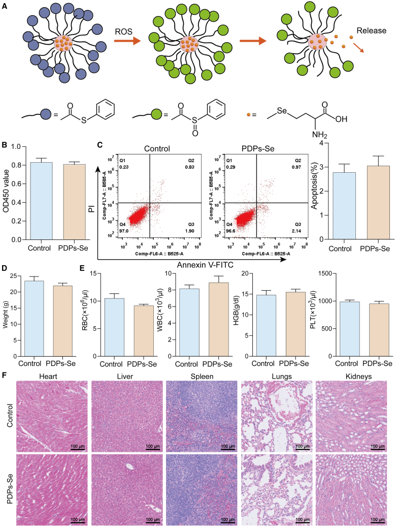
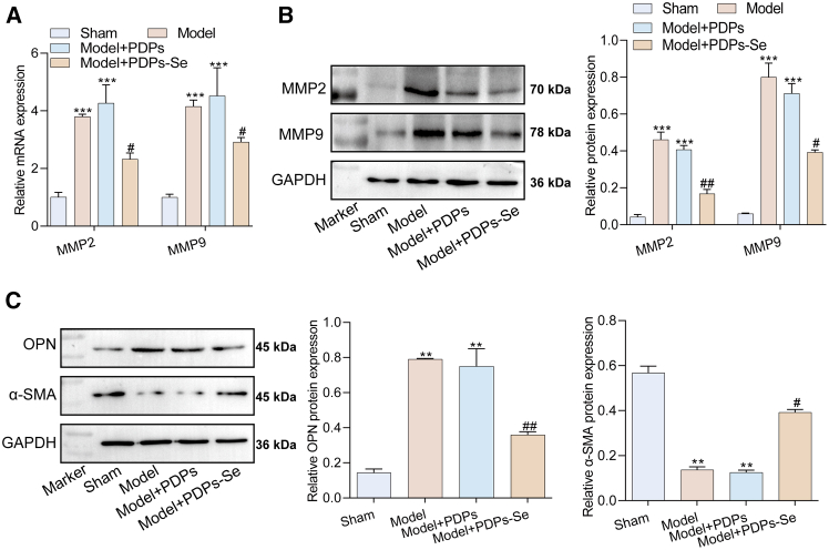
Oxidative stress, particularly ROS accumulation, plays a key role in the development of abdominal aortic aneurysm (AAA). Surgical treatments and current drugs for AAA have limitations, including lack of specificity and significant side effects. This study constructed ROS-responsive nanoparticles using phenylthio-modified dendritic polylysine (PDP) loaded with selenomethionine (PDPs-Se) for AAA treatment, and elucidated its mechanism of action. In-vitro studies revealed that PDPs-Se enhanced the clearance of ROS by increasing the levels of superoxide dismutase (SOD) and glutathione (GSH) while reducing malondialdehyde (MDA) levels. Furthermore, PDPs-Se upregulated the expression levels of GPX4, SLC7A11, and FTH1 to suppress ferroptosis and modulate the differentiation of vascular smooth muscle cells (VSMCs) from a synthetic to a contractile phenotype. In-vivo experiments revealed that PDPs-Se attenuated the progression of AAA by inhibiting oxidative stress responses and improving the aortic wall thickness, indicating its potential as an approach for AAA therapy.

Keywords: Bioengineering; Biological sciences; Nanoparticles; Therapeutics.


Conflict of interest statement

The authors declare that they have no competing interests.

Figures

None
Graphical abstract
Scheme 1
Scheme 1
Schematic illustration of the synthesis of PDPs-Se nanoparticles and their application to AAA treatment
Figure 1
Figure 1
Synthesis and characterization of PDPs and PDPs-Se nanoparticles (A) Synthesis reaction formula of PDPs-Se. (B) FTIR spectra of the dendritic polylysine and PDPs. (C) UV-vis spectra of dendritic polylysine and PDPs. (D) Photographs of the dendritic polylysine and PDPs dispersed in deionized water. (E) SEM images of PDPs-Se nanoparticles. (F) Particle size distribution of PDPs-Se. (G) Zeta potential of dendritic polylysine and PDPs dispersed in deionized water.
Figure 2
Figure 2
Biocompatibility of the PDPs-Se nanoparticles (A) Schematic illustration of encapsulation of selenocysteine in Se-containing nanoparticles. (B) Cell viability of VSMCs was assessed using the CCK8 assay. (C) Analysis of VSMCs apoptosis by flow cytometry. (D) Mouse weight measured using an electronic balance. (E) Detection of blood routine with a blood cell analyzer. (F) Evaluation of condition of mouse heart, liver, spleen, lung, and kidney through HE staining (scale bar: 100 μm). (n = 3).
Figure 3
Figure 3
PDPs-Se inhibited ferroptosis in ANG-II-induced VSMCs (A) The migration ability of the VSMCs was examined by Transwell experiment (scale bar: 200 μm). (B) Flow cytometry was used to evaluate the levels of ROS in VSMCs. (C) ELISA was used to evaluate the levels of SOD, MDA, and GSH in VSMCs. (D) Immunofluorescence analysis of GPX4 in VSMCs (scale bar: 200 μm). (E) Western blotting was conducted to detect the protein levels of FTH1 and SLC7A11 in VSMCs. n = 3, data are represented as mean ± SD. t test was utilized for statistical analysis. ∗∗p < 0.01, ∗∗∗p < 0.001 vs. Control group, #p < 0.05, ##p < 0.01vs ANG-II+PDPs group.
Figure 4
Figure 4
Inhibition of metabolic imbalance in ANG-II-induced VSMCs by PDPs-Se (A) The mRNA levels of MMP2 and MMP9 in VSMCs detected by RT-qPCR. (B) Protein levels of MMP2 and MMP9 in VSMCs detected by western blot. (C) The mRNA levels of OPN and α-SMA in VSMCs detected by RT-qPCR. (D) Immunofluorescence staining for OPN and α-SMA proteins in VSMCs (scale bar: 200 μm). n = 3, data are represented as mean ± SD. t test was utilized for statistical analysis. ∗∗p < 0.01, ∗∗∗p < 0.001 vs. Control group, #p < 0.05, ###p < 0.001vs ANG-II+PDPs group.
Figure 5
Figure 5
Inhibition of AAA procession in vivo by PDPs-Se (A) Photographs and diameter of abdominal aortas collected from different groups of treated mice. (B) HE staining of the abdominal aorta (scale bar: 200 μm). (C) TUNEL staining estimated the apoptosis levels of VSMCs (scale bar: 100 μm). n = 3, data are represented as mean ± SD. t test was utilized for statistical analysis. ∗∗p < 0.01 vs. sham group, ##p < 0.01 vs. Model+PDPs group.
Figure 6
Figure 6
Inhibition of ferroptosis in AAA mice by PDPs-Se (A) The levels of SOD, MDA, and GSH in VSMCs detected by ELISA. (B) The mRNA levels of GPX4, FTH1, and SLC7A11 in aneurysm tissue detected by RT-qPCR. (C) Immunofluorescence staining of GPX4 in aneurysm tissue. (D) Protein levels of FTH1 and SLC7A11 in aneurysm tissue detected by western blot. n = 3, data are represented as mean ± SD. t test was utilized for statistical analysis. ∗p < 0.05, ∗∗p < 0.01, ∗∗∗p < 0.01 vs. sham group, #p < 0.05, ##p < 0.01, ###p < 0.001 vs. Model+PDPs group.
Figure 7
Figure 7
Reversal of phenotypic transformation of AAA mice by PDPs-Se (A) The mRNA levels of MMP2 and MMP9 in aneurysm tissues detected by RT-qPCR. (B) Protein levels of MMP2 and MMP9 in aneurysm tissue detected by western blot. (C) The expression levels of OPN and α-SMA in aneurysm tissues detected by western blot. n = 3, data are represented as mean ± SD. t test was utilized for statistical analysis. ∗∗p < 0.01, ∗∗∗p < 0.001 vs. sham group, #p < 0.05, ##p < 0.01 vs. Model+PDPs group.

Similar articles

References

    1. Emeto T.I., Moxon J.V., Au M., Golledge J. Oxidative stress and abdominal aortic aneurysm: potential treatment targets. Clin. Sci. 2016;130:301–315. doi: 10.1042/cs20150547. - DOI - PubMed
    1. Sulistyowati E., Huang S.E., Cheng T.L., Chao Y.Y., Li C.Y., Chang C.W., Lin M.X., Lin M.C., Yeh J.L. Vasculoprotective Potential of Baicalein in Angiotensin II-Infused Abdominal Aortic Aneurysms through Inhibiting Inflammation and Oxidative Stress. Int. J. Mol. Sci. 2023;24 doi: 10.3390/ijms242116004. - DOI - PMC - PubMed
    1. Dyall-Smith D. Alopecia areata in a renal transplant recipient on cyclosporin. Australas. J. Dermatol. 1996;37:226–227. doi: 10.1111/j.1440-0960.1996.tb01066.x. - DOI - PubMed
    1. Gentille-Lorente D.I., Salvadó-Usach T. [Screening for abdominal aortic aneurysm by means of transthoracic echocardiography] Rev. Esp. Cardiol. 2011;64:395–400. doi: 10.1016/j.recesp.2010.11.009. - DOI - PubMed
    1. Golledge J., Moxon J.V., Singh T.P., Bown M.J., Mani K., Wanhainen A. Lack of an effective drug therapy for abdominal aortic aneurysm. J. Intern. Med. 2020;288:6–22. doi: 10.1111/joim.12958. - DOI - PubMed

LinkOut - more resources