. 2025 Feb 15;28(3):112033.
doi: 10.1016/j.isci.2025.112033. eCollection 2025 Mar 21.

Cell surface heparan sulfate is an attachment receptor for grass carp reovirus

Affiliations

Cell surface heparan sulfate is an attachment receptor for grass carp reovirus

Qian Wang et al. iScience. .

Abstract

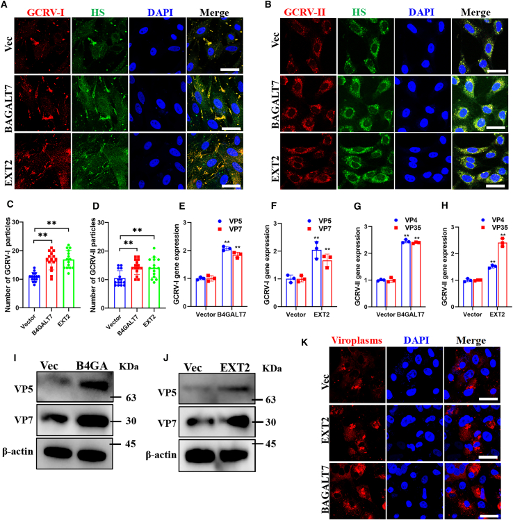
Grass carp reovirus (GCRV) causes hemorrhagic disease in grass carp, leading to significant economic losses in China's aquaculture. However, the cellular receptors responsible for the initiation of GCRV infection remain unclear. This study reveals that cell surface heparan sulfate (HS) acts as a crucial attachment receptor for GCRV. Removing HS with heparinase significantly reduces GCRV attachment and infection. Both HS and its homologue, heparin, inhibit the attachment of GCRV to cells. Altering HS levels in cells affects GCRV attachment and infection accordingly. GCRV outer capsid proteins VP5, VP56, and VP35, as well as purified GCRV virions, directly bind to HS. Pretreating GCRV with heparin or feeding grass carp with feed containing heparin significantly reduces mortality caused by GCRV infection. Collectively, these results highlight the crucial role of HS as an attachment receptor for GCRV and therefore provide a promising target for the prevention and control of this virus.

Keywords: Aquaculture; Aquaculture diseases; Biochemistry; Cell biology; Virology.


Conflict of interest statement

The authors declare no competing interests.

Figures

None
Graphical abstract
Figure 1
Figure 1
Removal of cell surface heparan sulfate significantly reduced GCRV attachment and infection See also Figure S1. (A and B) Immunofluorescence analysis of GCRV-I (A) and GCRV -II (B) attached cells that untreated or treated with heparinase I. Scale bar = 10 μm. (C and D) Quantitative analysis the number of GCRV-I (C) and GCRV-II (D) particles that attached to heparinase I treated or untreated cells. Data are represented as mean (n = 15) ± SD. ∗∗ indicates p < 0.01. (E and F) RT-qPCR analysis of GCRV-I (E) and GCRV -II (F) attached cells that mock treated or treated with heparinase I. Data are represented as mean (n = 3) ± SD. ∗ indicates p < 0.05, ∗∗ indicates p < 0.01. (G–I) Western blotting (G), cytopathic effect analysis (H) and plaque assay (I) of GCRV-I infected cells that untreated or treated with heparinase I. Scale bar = 100 μm.
Figure 2
Figure 2
Heparan sulfate and its homolog inhibits GCRV bind to cells (A and B) Immunofluorescence analysis of cells that attached with HS or heparin preincubated GCRV-I (A) or GCRV-II (B). Scale bar = 10 μm. (C and D) Quantitative analysis the effect of HS or heparin preincubation on the number of GCRV-I (C) and GCRV-II (D) particles that attached to cell surface. Data are represented as mean (n = 15) ± SD. ∗∗ indicates p < 0.01. (E–H) RT-qPCR analysis of cells that attached with HS or heparin preincubated GCRV-I (E, F) or GCRV-II (G, H). Data are represented as mean (n = 3) ± SD. ∗∗ indicates p < 0.01. (I–M) Western blotting (I, J), plaque assay (K, L), and cytopathic effect analysis (M) of cells that infected with HS or heparin preincubated GCRV-I. Scale bar = 100 μm.
Figure 3
Figure 3
Heparan sulfate-knockdown cells impact GCRV attachment and infection See also Figure S1. (A and B) Immunofluorescence analysis of control or B4GALT7-knockdown cells that attached with GCRV-I (A) or GCRV-II (B). Scale bar = 10 μm. (C and D) Quantitative analysis the number of GCRV-I (C) and GCRV-II (D) particles that attached to control or B4GALT7-knockdown cells. Data are represented as mean (n = 15) ± SD. ∗∗ indicates p < 0.01. (E–H) RT-qPCR analysis of control or heparan sulfate-knockdown cells that attached with GCRV-I (E, F) or GCRV-II (G, H). (I and J) Western blotting analysis of control or heparan sulfate-knockdown cells that infected with GCRV-I. (K and L) Immunofluorescence analysis of the viroplasms in mock or heparan sulfate-knockdown cells that infected with GCRV-I. Scale bar = 10 μm.
Figure 4
Figure 4
Elevation of heparan sulfate biosynthesis facilitates GCRV attachment and infection See also Figure S1. (A and B) Immunofluorescence analysis of mock or heparan sulfate biosynthesis elevated cells that attached with GCRV-I (A) or GCRV-II (B). Scale bar = 10 μm. (C and D) Quantitative analysis the number of GCRV-I (C) and GCRV-II (D) particles that attached to control or heparan sulfate biosynthesis elevated cells. Data are represented as mean (n = 15) ± SD. ∗∗ indicates p < 0.01. (E–H) RT-qPCR analysis of control or heparan sulfate biosynthesis elevated cells that attached with GCRV-I (E, F) or GCRV-II (G, H). (I and J) Western blotting analysis of control or heparan sulfate biosynthesis elevated cells that infected with GCRV-I. (K) Immunofluorescence analysis of the viroplasms in mock or heparan sulfate biosynthesis elevated that infected with GCRV-I. Scale bar = 10 μm.
Figure 5
Figure 5
GCRV capsid proteins and purified GCRV virions bind to heparan sulfate See also Figures S2 and S3. (A and B) The direct binding between GCRV capsid proteins and HS-conjugated Sepharose beads. (C and D) Heparan sulfate blocked the binding between GCRV capsid proteins and HS-conjugated Sepharose beads. (E and F) The direct binding between purified GCRV virions and HS-conjugated Sepharose beads. (G and H) Chondroitin sulfate (CS) did not affect the binding between GCRV capsid proteins and HS. The fractions of input (Input), supernatant after incubation (S), the fifth wash solution (W5), and the eluate solution (eluate) were detected by western blotting with the antibodies against the corresponding proteins.
Figure 6
Figure 6
Pretreatment of GCRV with heparin reduce the mortality of grass carp after GCRV infection See also Figure S4. (A) Percent of survival in four different grass carp groups. (B) The hemorrhagic symptoms of fish from the group injected with GCRV-II suspension or injected with GCRV-II suspension preincubated with heparin. Scale bar = 1 cm (C and D) RT-qPCR analysis the relative copy number of GCRV-II in liver (C) and intestine (D) of different grass carp groups. Data are represented as mean (n = 3) ± SD. ∗∗ indicates p < 0.01. (E) Histological section analysis of liver and intestine of different grass carp groups. Scale bar = 20 μm. (F) TEM analysis of kidney samples from different grass carp groups. The red frames in the upper panes were enlarged in the bottom panes. Scale bar = 2 μm and 1μm.
Figure 7
Figure 7
Feeding grass carp with feed containing heparin decrease the mortality of grass carp after GCRV infection See also Figure S5. (A) Percent of survival in five different grass carp groups. (B) The hemorrhagic symptoms in GCRV-infected fish that fed with normal feed or fed with feed containing heparin. Scale bar = 1 cm (C and D) RT-qPCR analysis the relative copy number of GCRV-II in liver (C) and intestine (D) of five different grass carp groups. Data are represented as mean (n = 3) ± SD. ∗∗ indicates p < 0.01. (E) Histological section analysis of liver and intestine of five different grass carp groups. Scale bars = 20 μm. (F) TEM analysis of kidney samples from five different grass carp groups. The red frames in the upper panes were enlarged in the bottom panes. Scale bar = 2 μm and 1μm.

Similar articles

References

    1. Fisheries Bureau of Ministry of Agriculture and Rural Affairs in China . China Agriculture Press; 2023. China Fishery Statistical Yearbook of 2022.
    1. Wang Q., Zeng W., Liu C., Zhang C., Wang Y., Shi C., Wu S. Complete genome sequence of a reovirus isolated from grass carp, indicating different genotypes of GCRV in China. J. Virol. 2012;86 - PMC - PubMed
    1. Fang Q., Attoui H., Cantaloube J.F., Biagini P., Zhu Z., de Micco P., de Lamballerie X. Sequence of genome segments 1, 2, and 3 of the grass carp reovirus (Genus Aquareovirus, family Reoviridae) Biochem. Biophys. Res. Commun. 2000;274:762–766. - PubMed
    1. Ye X., Tian Y.Y., Deng G.C., Chi Y.Y., Jiang X.Y. Complete genomic sequence of a reovirus isolated from grass carp in China. Virus Res. 2012;163:275–283. - PubMed
    1. Pei C., Ke F., Chen Z.Y., Zhang Q.Y. Complete genome sequence and comparative analysis of grass carp reovirus strain 109 (GCReV-109) with other grass carp reovirus strains reveals no significant correlation with regional distribution. Arch. Virol. 2014;159:2435–2440. - PubMed

LinkOut - more resources