

A Hallux Valgus Surgical Planning Survey Using WBCT-based 3D Printing
- PMID: 40104096
- PMCID: PMC11915313
- DOI: 10.1177/24730114251325854
A Hallux Valgus Surgical Planning Survey Using WBCT-based 3D Printing
Abstract
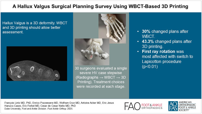
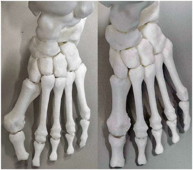
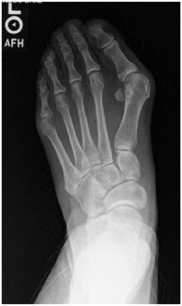
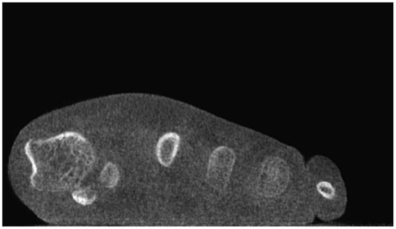
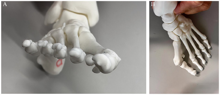
Background: Recent literature highlights the importance of treating hallux valgus (HV) as a 3-dimensional (3D) deformity. Although 3D printing may enhance visualization of the multiplanar aspects of HV, its influence on surgical planning remains unclear. This study assessed changes in surgical plans when surgeons sequentially reviewed 2D radiographs, 3D weightbearing computed tomography (WBCT), and 3D-printed models, hypothesizing that 3D printing would have the greatest impact.
Methods: A single HV case (a 40-year-old woman, intermetatarsal angle [IMA] 21 degrees, HV angle [HVA] 47 degrees) was evaluated by 30 surgeons in a masked, stepwise manner. Surgical plans were recorded at each stage. Surgeons rated the influence of WBCT and 3D printing using a 5-point Likert scale. A follow-up survey examined the effect of these technologies on correction amplitudes.
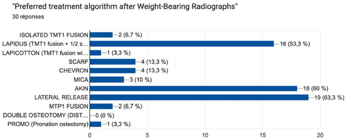
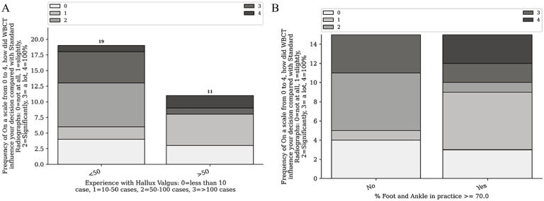
Results: The participants were mostly early career surgeons (median age 35.5 years, 2 years in practice). WBCT was accessible to 43.3% and used in 30% of HV cases, whereas 3D printing was accessible to 23.3% and used in 6.6%. Changes in the treatment algorithm occurred in 30% of cases after WBCT and in 43.3% after 3D printing. Significant differences (P < .05) were observed for the Lapicotton procedure between radiography and WBCT, and between WBCT and 3D printing. Surgeons performing <50 HV cases annually or with >70% Foot and Ankle specialization were more influenced by WBCT. Follow-up data (n = 23) indicated that WBCT and 3D printing influenced correction amplitudes, particularly for pronation and distal metatarsal articular angle (DMAA), more than for the IMA.
Discussion: Both WBCT and 3D printing influenced surgical planning, mostly explained by changes in first ray tarsometatarsal procedures. The rotational components (pronation and DMAA) were perceived as the most significantly affected. Future studies should explore cost-effectiveness, patient outcomes, and the utility of combining WBCT and 3D printing in other deformities requiring multiplanar corrections.Level of Evidence: Level IV, cross-sectional survey.
Keywords: 3D-printing; cone beam; hallux valgus; weightbearing computed tomography.
© The Author(s) 2025.
Conflict of interest statement
The author(s) declared the following potential conflicts of interest with respect to the research, authorship, and/or publication of this article: François Lintz, MD, PhD, reports disclosures from Paragon28 (consultant, shareholder), CurvebeamAI (consultant, shareholder), Newclip Technics (consultant, royalties), Podonov (consultant, royalties), LINNOV (founder, shareholder), Followinvest (shareholder), and International WBCT Society (cofounder, past president). Antoine Acker, MD, reports disclosures from CurvebeamAI (shareholder). Eric Ferkel, MD, reports disclosures from Ossio, Arthrex, Smith & Nephew, Medartis (consulting fees, royalties), International Bone Research Association (IBRA), American Academy of Orthopaedic Surgeons (AAOS) (support for attending meetings), and AAOS, American Orthopaedic Society for Sports Medicine (AOSSM), American Orthopaedic Foot & Ankle Society (AOFAS), Arthroscopy Association of North America (AANA) leadership roles. Cesar de Cesar Netto, MD, PhD, reports disclosures from Paragon28 (consultant, medical advisory board, royalties), CurvebeamAI (consultant, shareholder), Ossio (consultant), Zimmer (consultant), Stryker (consultant), International WBCT Society (cofounder, President), Exactech (consultant), Arthrex (consultant), Tayco Brace (shareholder), Extremity Medical (consultant), AOFAS committee member, and Foot Ankle Clinics (editor in chief). Disclosure forms for all authors are available online.
Figures
Similar articles
-
The Role of Weightbearing Computed Tomography Scan in Hallux Valgus.Foot Ankle Int. 2021 Mar;42(3):287-293. doi: 10.1177/1071100720962398. Epub 2020 Nov 4. Foot Ankle Int. 2021. PMID: 33148045
-
Distal Metatarsal Articular Angle in Hallux Valgus Deformity. Fact or Fiction? A 3-Dimensional Weightbearing CT Assessment.Foot Ankle Int. 2022 Apr;43(4):495-503. doi: 10.1177/10711007211051642. Epub 2021 Nov 13. Foot Ankle Int. 2022. PMID: 34779306
-
A Comprehensive Weightbearing Computed Tomography Study on Patients With Hallux Valgus: Exploring Multiplanar Deformity Interrelationships.Foot Ankle Int. 2025 Mar;46(3):343-356. doi: 10.1177/10711007241309912. Epub 2025 Jan 24. Foot Ankle Int. 2025. PMID: 39861960
-
Weightbearing Imaging Assessment of Midfoot Instability in Patients with Confirmed Hallux Valgus Deformity: A Systematic Review of the Literature.Diagnostics (Basel). 2024 Jan 16;14(2):193. doi: 10.3390/diagnostics14020193. Diagnostics (Basel). 2024. PMID: 38248070 Free PMC article. Review.
-
Modified Lindgren-Turan Osteotomy for Hallux Valgus Deformity - a Review of 60 Cases.Acta Chir Orthop Traumatol Cech. 2018;85(5):325-330. Acta Chir Orthop Traumatol Cech. 2018. PMID: 30383528 Review. English.
References
-
- 3D-Printing Surgical Planning Study. https://forms.gle/1DpH9J6MFbpUprLU8
-
- 3D-Printing Surgical Planning Survey. https://forms.gle/Tk1KAvaQgpTpq1Hg6
LinkOut - more resources
Full Text Sources