

Creating anatomically derived, standardized, customizable, and three-dimensional printable head caps for functional neuroimaging
- PMID: 40104430
- PMCID: PMC11915464
- DOI: 10.1117/1.NPh.12.1.015016
Creating anatomically derived, standardized, customizable, and three-dimensional printable head caps for functional neuroimaging
Abstract
Significance: Consistent and accurate probe placement is a crucial step toward enhancing the reproducibility of longitudinal and group-based functional neuroimaging studies. Although the selection of headgear is central to these efforts, there does not currently exist a standardized design that can accommodate diverse probe configurations and experimental procedures.
Aim: We aim to provide the community with an open-source software pipeline for conveniently creating low-cost, three-dimensional (3D) printable neuroimaging head caps with anatomically significant landmarks integrated into the structure of the cap.
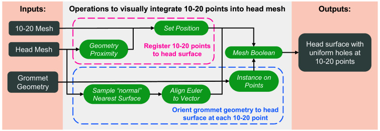
Approach: We utilize our advanced 3D head mesh generation toolbox and 10-20 head landmark calculations to quickly convert a subject's anatomical scan or an atlas into a 3D printable head cap model. The 3D modeling environment of the open-source Blender platform permits advanced mesh processing features to customize the cap. The design process is streamlined into a Blender add-on named "NeuroCaptain."
Results: Using the intuitive user interface, we create various head cap models using brain atlases and share those with the community. The resulting mesh-based head cap designs are readily 3D printable using off-the-shelf printers and filaments while accurately preserving the head geometry and landmarks.
Conclusions: The methods developed in this work result in a widely accessible tool for community members to design, customize, and fabricate caps that incorporate anatomically derived landmarks. This not only permits personalized head cap designs to achieve improved accuracy but also offers an open platform for the community to propose standardizable head caps to facilitate multi-centered data collection and sharing.
Keywords: 10-20 system; 3D printing; electroencephalography; functional near-infrared spectroscopy; head cap; mesh generation; personalized medicine.
© 2025 The Authors.
Figures


Update of
-
Creating anatomically-derived, standardized, customizable, and three-dimensional printable head caps for functional neuroimaging.bioRxiv [Preprint]. 2024 Dec 17:2024.08.30.610386. doi: 10.1101/2024.08.30.610386. bioRxiv. 2024. Update in: Neurophotonics. 2025 Jan;12(1):015016. doi: 10.1117/1.NPh.12.1.015016. PMID: 39257741 Free PMC article. Updated. Preprint.
Similar articles
-
Creating anatomically-derived, standardized, customizable, and three-dimensional printable head caps for functional neuroimaging.bioRxiv [Preprint]. 2024 Dec 17:2024.08.30.610386. doi: 10.1101/2024.08.30.610386. bioRxiv. 2024. Update in: Neurophotonics. 2025 Jan;12(1):015016. doi: 10.1117/1.NPh.12.1.015016. PMID: 39257741 Free PMC article. Updated. Preprint.
-
ninjaCap: a fully customizable and 3D printable headgear for functional near-infrared spectroscopy and electroencephalography brain imaging.Neurophotonics. 2024 Jul;11(3):036601. doi: 10.1117/1.NPh.11.3.036601. Epub 2024 Aug 27. Neurophotonics. 2024. PMID: 39193445 Free PMC article.
-
ninjaCap: A fully customizable and 3D printable headgear for fNIRS and EEG brain imaging.bioRxiv [Preprint]. 2024 May 16:2024.05.14.594159. doi: 10.1101/2024.05.14.594159. bioRxiv. 2024. Update in: Neurophotonics. 2024 Jul;11(3):036601. doi: 10.1117/1.NPh.11.3.036601. PMID: 38798389 Free PMC article. Updated. Preprint.
-
Spatial registration for functional near-infrared spectroscopy: from channel position on the scalp to cortical location in individual and group analyses.Neuroimage. 2014 Jan 15;85 Pt 1:92-103. doi: 10.1016/j.neuroimage.2013.07.025. Epub 2013 Jul 25. Neuroimage. 2014. PMID: 23891905 Review.
-
Materials Properties of Printable Edible Inks and Printing Parameters Optimization during 3D Printing: a review.Crit Rev Food Sci Nutr. 2019;59(19):3074-3081. doi: 10.1080/10408398.2018.1481823. Epub 2018 Jun 20. Crit Rev Food Sci Nutr. 2019. PMID: 29856675 Review.
References
-
- Quaresima V., “Functional near-infrared spectroscopy (fNIRS) for assessing cerebral cortex function during human behavior in natural/social situations: a concise review - Valentina Quaresima, Marco Ferrari, 2019,” Organ. Res. Methods 22(1), 46–68 (2019).10.1177/1094428116658959 - DOI
Grants and funding
LinkOut - more resources
Full Text Sources
Miscellaneous
