

Immunohistochemical Expression of DAPK-1 in Oral Leukoplakia And Oral Squamous Cell Carcinoma: A Preliminary Study
- PMID: 40104466
- PMCID: PMC11916529
- DOI: 10.7759/cureus.79085
Immunohistochemical Expression of DAPK-1 in Oral Leukoplakia And Oral Squamous Cell Carcinoma: A Preliminary Study
Abstract
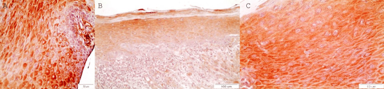
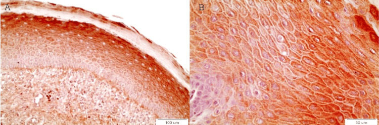
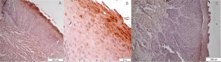
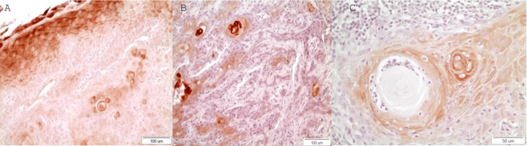
Introduction: The silencing of death-associated protein kinase 1 (DAPK-1) is an effective way of inactivating a tumor-suppressing mechanism. The aim of this study was to investigate the immunohistochemical expression of DAPK-1 in oral leukoplakia (OL) and oral squamous cell carcinoma (OSCC).
Methods: The immunohistochemical (IHC) detection of DAPK-1 was carried out in cases of OLs and OSCCs. DAPK-1 molecules' tissue distribution in OLs/OSCCs tissues was evaluated using semiquantitative immunohistochemistry in representative paraffin-embedded tissue samples (57 in total) from 2004-2019, retrieved from the archives of the Department of Oral Medicine/Pathology, School of Dentistry, Aristotle University of Thessaloniki, Greece and the St Lukas Hospital of Thessaloniki, Greece. The inclusion criterion was the presence of sufficient precancerous or cancerous biological material (estimated as more than 70% per tissue specimen) in the paraffin cubes. The exclusion criterion was the opposite, i.e. the lack of sufficient material due to previous sections. Statistics for IHC were evaluated by a non-parametric Mann-Whitney U Test. A two-sided p-value < 0.05 was considered statistically significant.
Results: DAPK-1 IHC expression was increased in OLs without dysplasia and with OLs with mild dysplasia compared to moderate/severe dysplasia (p=0.019, Mann-Whitney U Test) and OSCCs (p=0.003, Mann-Whitney U Test). Conclusions: DAPK-1 seemed to function as an oncosuppressor molecular biomarker, as its expression was decreased in areas of cellular dysplasia in OLs and in areas of OSCCs composed of less differentiated cells. The clinical application of this biomarker is that the positively stained, potentially malignant lesions are less likely to transition into malignancy, and cancerous lesions are more likely to behave non-aggressively. On the other hand, the lack of staining could signify the loss of this oncosuppressing ability, and it could be a potential prognostic biomarker for OSCC's aggressive biologic behavior if considered with other clinical parameters and a prognostic factor of malignant transformation of potentially malignant lesions. Since this is a preliminary study, more studies with larger sample sizes are required to support these conclusions.
Keywords: cancer biomarkers; cancer stem cells; dapk-1; immunohistochemistry ihc; oral leukoplakia; oral squamous cell carcinoma.
Copyright © 2025, Papadopoulos et al.
Conflict of interest statement
Human subjects: Consent for treatment and open access publication was obtained or waived by all participants in this study. Ethics Committee of the School of Dentistry, Aristotle University of Thessaloniki, Greece issued approval 29/21.11.2018. Animal subjects: All authors have confirmed that this study did not involve animal subjects or tissue. Conflicts of interest: In compliance with the ICMJE uniform disclosure form, all authors declare the following: Payment/services info: All authors have declared that no financial support was received from any organization for the submitted work. Financial relationships: All authors have declared that they have no financial relationships at present or within the previous three years with any organizations that might have an interest in the submitted work. Other relationships: All authors have declared that there are no other relationships or activities that could appear to have influenced the submitted work.
Figures
References
-
- PD-1/PD-L1, Treg-related proteins, and tumour-infiltrating lymphocytes are associated with the development of oral squamous cell carcinoma. Kujan O, Agag M, Smaga M, Vaishnaw Y, Idrees M, Shearston K, Farah CS. Pathology. 2022;54:409–416. - PubMed
-
- Survival predictors and outcomes of patients with recurrent and/or metastatic head and neck cancer treated with chemotherapy plus cetuximab as first-line therapy: A real-world retrospective study. Pontes F, Garcia AR, Domingues I, et al. Cancer Treat Res Commun. 2021;27:100375. - PubMed
LinkOut - more resources
Full Text Sources