Review
. 2025 Mar 4:12:1546719.
doi: 10.3389/fvets.2025.1546719. eCollection 2025.

How facial expressions reveal acute pain in domestic animals with facial pain scales as a diagnostic tool

Affiliations
Review

How facial expressions reveal acute pain in domestic animals with facial pain scales as a diagnostic tool

Daniel Mota-Rojas et al. Front Vet Sci. .

Abstract

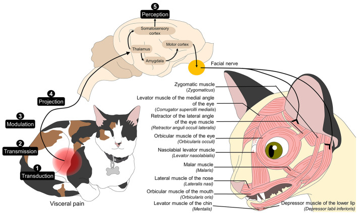
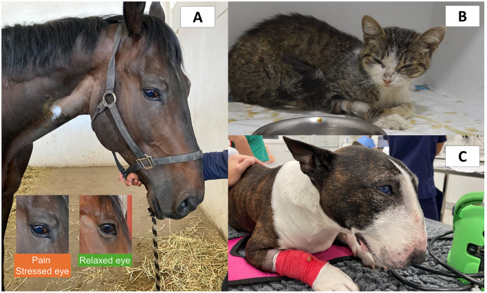
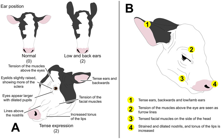
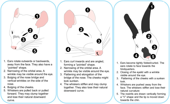
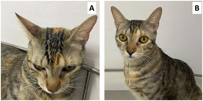
The growing interest in managing and recognizing pain in animals has led to the search for more sensitive methods to evaluate it, especially because some species conceal any visible changes associated with pain or are not easily assessed. Research has shown that an animal's facial expression changes when exposed to painful stimuli. Thus, developing several pain scales (grimace scales) in species such as horses, cattle, pigs, sheep, donkeys, rabbits, rats, mice, and cats has helped to improve the study of pain in veterinary medicine. The possibility of using facial expression as an indicator of pain is due to the direct relationship between the activation of different regions of the Central Nervous System such as the somatosensory cortex, prefrontal cortex, amygdala, hippocampus, and hypothalamus, and their connections with the motor cortex to elicit motor responses including the movement of facial muscles. The present review aims to discuss the neurobiological association between acute pain and facial expressions in animals. It will analyze the importance of facial expression characterization and the use of grimace scales in farm, companion, and laboratory species.

Keywords: acute pain; facial expressions; grimace; neurobiology; pain.


Conflict of interest statement

The authors declare that the research was conducted in the absence of any commercial or financial relationships that could be construed as a potential conflict of interest. The author(s) declared that they were an editorial board member of Frontiers, at the time of submission. This had no impact on the peer review process and the final decision.

Figures

Figure 1
Figure 1
Association between pain neurobiology and facial expression. Noxious stimuli are transduced through free nerve endings. The nociceptive signals are transmitted by peripheral nerve fibers and modulated and projected from the dorsal horn of the spinal cord to various regions of the central nervous system. After reaching cerebral structures such as the thalamus, its connections to the amygdala and, consequently, to the motor cortex star the efferent motor response to pain. The motor cortex directly connects to the facial nucleus in the pons and, in turn, to the cranial nerve VII, which innervates the facial muscles. Activating these regions leads to the production of facial muscle movement that generate facial expressions that can be associated with the perception of pain.
Figure 2
Figure 2
Comparison of facial expression associated with pain in different species. (A) A male quarter horse with traumatic laminitis. Changes in facial expression such as tension of the upper orbital muscles, tension of the lower lip, dilation of the nostrils, and tension of the facial muscles can be observed. The lower rectangles emphasize the changes observed in a horse with pain. When frightened, anxious, and in pain, the extraorbital eyelid muscles tend to hold more tension and create a “beak” over the eye. In contrast, the upper eyelid of the eye pulls up in a triangular shape due to muscle tension of the levator muscle of the medial angle of the eye (m. levator anguli oculi medialis). (B) Five-year-old male cat with acute pain due to interstitial cystitis. Changes in facial expression can be seen such as tension of the orbital muscles, head with a low position, tension in the whisker muscles, and slight flattening of the ears to the side. (C) Six-year-old Bull Terrier dog with acute pain due to pericardial effusion. In this picture, the tension of the orbital muscles, low head position, the tension of the facial muscles, retraction of the corner of the lip, and flattening of the ears can be observed. Images provided by the Equine Hospital of the Faculty of Higher Studies Cuautitlán, and to the Specialty Hospital of the Catholic University of Valencia.
Figure 3
Figure 3
Facial expression related to pain in cattle and calves. (A) The Cow Pain Scale. Although this behavioral-based assessment method does not focus on facial expression, a tense facial expression and changes in ear position are included in the evaluation of pain. Numbers inside the parenthesis represent the pain score. (B) Calf Grimace Scale and the main FAU.
Figure 4
Figure 4
Main changes observed in the facial expression of mice, rats, and rabbits when perceiving severe pain.
Figure 5
Figure 5
Four-year-old female domestic cat diagnosed with acute pancreatitis. (A) shows the flattening of the ears and the position of the whiskers forward, as well as the squinting of the eyelids, these being grimaces suggestive of moderate pain according to the FGS. In (B) you can see the same patient without pain after the administration of analgesics.

Similar articles

Cited by

References

    1. Broom DM. Welfare, stress, and the evolution of feelings. Adv Study Behav. (1998) 27:371–403. 10.1016/S0065-3454(08)60369-1 - DOI
    1. Cohen S, Beths T. Grimace scores: tools to support the identification of pain in mammals used in research. Animals. (2020) 10:1726. 10.3390/ani10101726 - DOI - PMC - PubMed
    1. Descovich KA, Wathan J, Leach MC, Buchanan-Smith HM, Flecknell P, Farningham D, et al. . Facial expression: an under-utilised tool for the assessment of welfare in mammals. ALTEX. (2017) 34:409–29. 10.14573/altex.1607161 - DOI - PubMed
    1. Turner P V, Pang DS, Lofgren JL, A. Review of pain assessment methods in laboratory rodents. Comp Med. (2019) 69:451–67. 10.30802/AALAS-CM-19-000042 - DOI - PMC - PubMed
    1. McLennan K, Mahmoud M. Development of an automated pain facial expression detection system for sheep (ovis aries). Animals. (2019) 9:196. 10.3390/ani9040196 - DOI - PMC - PubMed

LinkOut - more resources