

A Case of Glomus Tumor of the Anterior Neck
- PMID: 40104558
- PMCID: PMC11912979
- DOI: 10.53045/jprs.2023-0027
A Case of Glomus Tumor of the Anterior Neck
Abstract
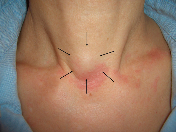
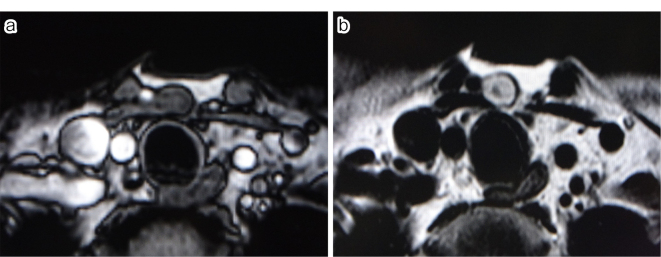
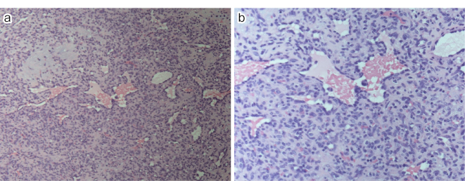
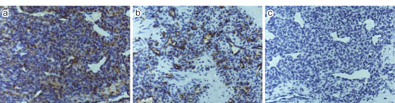
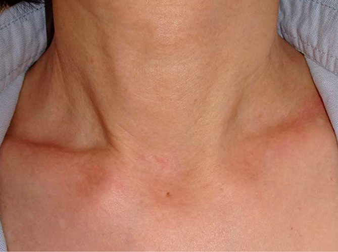
A 54-year-old woman presented with an anterior neck subcutaneous tumor that had appeared one month prior. Mild tenderness was noted. As a diagnosis was difficult to make based on physical examination and ultrasonography, a magnetic resonance imaging (MRI) scan was performed. Both examinations revealed a 1-cm subcutaneous mass with well-defined margins; the MRI scan was hypointense on T1-weighted images and slightly hyperintense with low point foci on T2-weighted images. Subsequently, an excisional biopsy was performed, and the pathologic diagnosis of glomus tumor was obtained. Glomus tumors usually present as a painful subcutaneous mass beneath the nail bed but may be painless or occur in areas other than the fingers. Because glomus tumors in the neck resemble a variety of diseases, their diagnosis may be delayed. This case highlights the importance of considering glomus tumors as a potential cause of neck subcutaneous tumors.
Keywords: anterior neck; diagnosis; glomus tumor; subcutaneous tumor; tenderness.
© 2024 The Japan Society of Plastic and Reconstructive Surgery.
Conflict of interest statement
Conflicts of Interest: There are no conflicts of interest.
Figures
Similar articles
-
Subungual glomus tumour: magnetic resonance imaging findings.Australas Radiol. 2007 Oct;51 Spec No.:B107-9. doi: 10.1111/j.1440-1673.2007.01797.x. Australas Radiol. 2007. PMID: 17875128 Review.
-
Subungual glomus tumor diagnosis based on imaging.J Dermatol. 2006 Jun;33(6):389-93. doi: 10.1111/j.1346-8138.2006.00092.x. J Dermatol. 2006. PMID: 16700827
-
Subungual onychomycosis due to Aspergillus niger mimicking a glomus tumor: A case report.Biomed Rep. 2017 Dec;7(6):532-534. doi: 10.3892/br.2017.994. Epub 2017 Sep 29. Biomed Rep. 2017. PMID: 29188057 Free PMC article.
-
Intravascular glomus tumor of the forearm causing chronic pain and focal tenderness.Case Rep Orthop. 2014;2014:619490. doi: 10.1155/2014/619490. Epub 2014 Feb 3. Case Rep Orthop. 2014. PMID: 24624306 Free PMC article.
-
Glomus tumor of the plantar arch: a case report with magnetic resonance imaging findings.Foot Ankle Int. 1997 Oct;18(10):672-4. doi: 10.1177/107110079701801014. Foot Ankle Int. 1997. PMID: 9347308 Review.
References
-
- Rimington TR, Lefton CS. Simultaneous solitary glomus tumors in nonadjacent digits. Am J Orthop. 2009 May;38:E82-4. - PubMed
Publication types
LinkOut - more resources
Full Text Sources
Research Materials