Review
. 2025 Feb 25:31:101614.
doi: 10.1016/j.mtbio.2025.101614. eCollection 2025 Apr.

Recent advances in bioactive hydrogel microspheres: Material engineering strategies and biomedical prospects

Affiliations
Review

Recent advances in bioactive hydrogel microspheres: Material engineering strategies and biomedical prospects

Junjiang Yue et al. Mater Today Bio. .

Abstract

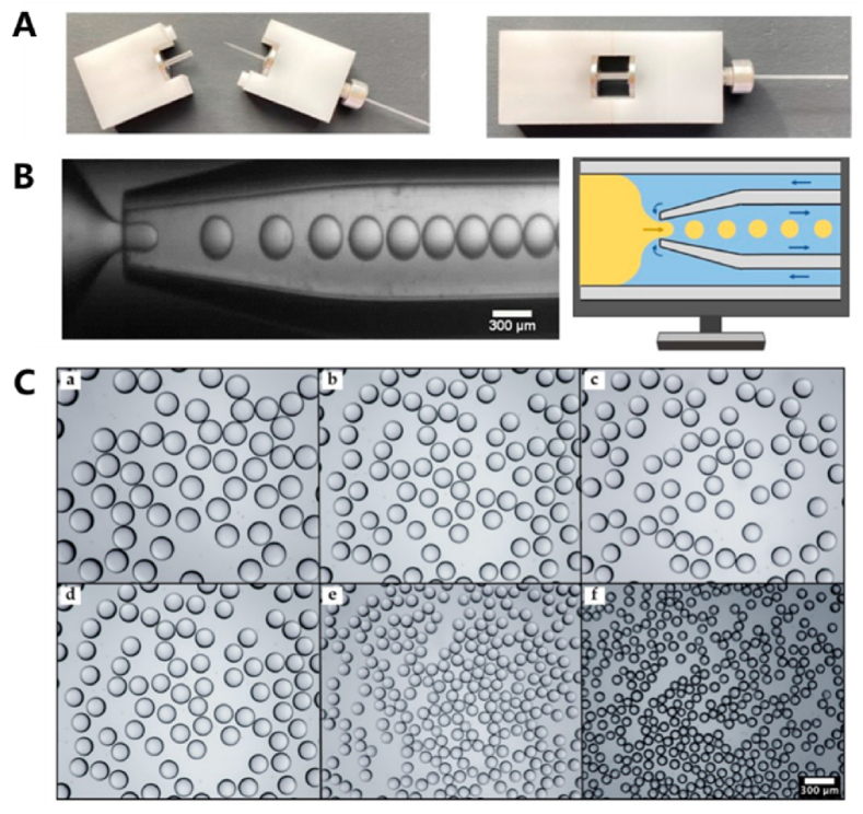
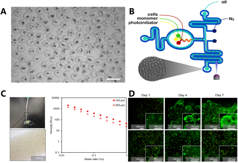
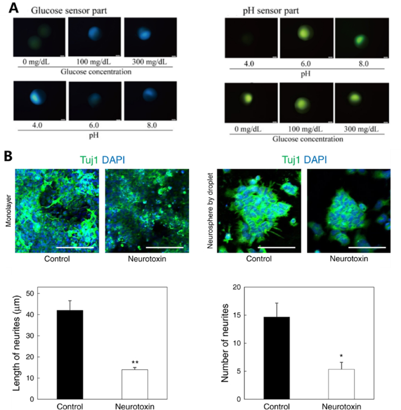
Hydrogel microspheres are a class of hydrophilic polymeric particles in microscale, which has been developed as a new type of functional biomaterials for wide-range biomedical applications in recent years. This review provides a comprehensive overview of the preparation methods for hydrogel microspheres, including droplet microfluidics, electrospray and emulsion was first summarized. At the same time, we analyze the impacts of these methods on the properties of hydrogel microspheres and explore various functionalization strategies for enhancing their bioactivity and expanding their biomedical applications. In addition, we discuss the recent advances and the further prospect of hydrogel microspheres in life science applications, particularly in cell biology research, bioanalysis and detection, as well as tissue repair and regeneration. By synthesizing the latest developments, this review aims to offer valuable insights and strategies for optimizing hydrogel microspheres in diverse application scenarios and inspire future research and practical innovations.

Keywords: 3D cell culture; Biofunctionalization; Biosensing; Hydrogel microspheres; Microfluidics; Tissue regeneration.


Conflict of interest statement

The authors declare that they have no known competing financial interests or personal relationships that could have appeared to influence the work reported in this paper.

Figures

Image 1
Graphical abstract
Scheme 1
Scheme 1
The preparations, biofunctionalizations and applications of hydrogel microspheres.
Fig. 1
Fig. 1
(A) The fabrication process of PDMS microfluidic chips: (a) Pouring PDMS onto the mold and allowing it to cross-link; (b) Peeling off the PDMS layer; (c) Puncturing fluid access holes; (d) Bonding the PDMS to a glass substrate and filling with fluorescein to enhance contrast. (B) A four-channel microfluidic chip device enables precise fluidic control. (C) Three-dimensional core-shell scaffolds facilitate the spatial organization and assembly of diverse cell types [39]. Copyright 2022, The Authors.
Fig. 2
Fig. 2
(A) A glass capillary device assembled from LEGO bricks. (B) The principle and schematic diagram of droplet formation process at the microscopic scale. (C) PEGDA particles generated by utilizing the inner capillary with diverse orifice diameters: (a) 400 μm; (b) 350 μm; (c) 300 μm; (d) 250 μm; (e) 200 μm; and (f) 150 μm. Reproduced with permission [43]. Copyright 2022, MDPI.
Fig. 3
Fig. 3
(A) A microfluidic manufacturing device for a syringe with coaxial placement. (B) The MCP membrane is like a sieve. The dispersed phase is the material that needs to pass through the sieve (the membrane). The pressure created by blocking the ports is similar to the force you would apply to push the material through the sieve. The continuous phase in the lower channel is like the container that catches the filtered material and combines with it to form the final product, which is the emulsion. Use a container to collect droplets from port b for further processing. (C) Fabricated silicon wafer microchannel reactor. Reproduced with permission [[47], [48], [49]]. Copyright 2020, Elsevier B.V. Copyright 2017, Elsevier B.V. Copyright 2023, American Chemical Society.
Fig. 4
Fig. 4
(A) Electrospray patterns and images showing particle formation under different voltages. (B) Diagram of preparation of microspheres by emulsion method. (C) Preparation of glucose and PH dual response Janus microspheres by UV-assisted centrifugation. (D) Schematic diagram of preparation of multistage hydrogels with ordered micro-nano structures. Reproduced with permission [51,52,54,57]. Copyright 2022, Elsevier B.V. Copyright 2024, American Chemical Society. Copyright 2021, MDPI. Copyright 2021, Science Partner Journals.
Fig. 5
Fig. 5
(A) Encapsulation of mESCs with PEG hydrogel microspheres. (B) Schematic diagram of a two-layer nitrogen jacket microfluidic device. (C) The viscosity of the hydrogel derived from tilapia decreases with the increase of shear rate. (D) Live/dead fluorescence was performed on 3D cell cultures of L929 cells in dECM particle hydrogel assemblies and dECM bulk gel for 7 days. Reproduced with permission [[69], [70], [71]]. Copyright 2016, Elsevier B.V. Copyright 2016, American Chemical Society. Copyright 2024, American Chemical Society.
Fig. 6
Fig. 6
(A) Formation of U-CMLV hydrogel microspheres (B) Alginate saline gelatin crosslinking process diagram and prepared hydrogel microspheres (C) Single-layer hydrogel microcapsules in simulated gastric fluid and simulated intestinal fluid for drug release (D) Core-shell hydrogel microcapsules in simulated gastric fluid and simulated intestinal fluid for drug release. Reproduced with permission [84,85]. Copyright 2023, John Wiley and Sons. Copyright 2024, Elsevier B.V.
Fig. 7
Fig. 7
(A) Fluorescence images of glucose and pH probe components at various glucose concentrations and pH conditions. (B) Drug screening analysis for neurospheres produced by droplet-based microfluidic devices: Images obtained through confocal laser scanning microscopy of monolayer neurons and quantitative analysis regarding the length and number of neurites originating from the neurospheres. Reproduced with permission [57,87]. Copyright 2021, MDPI. Copyright 2020, Springer Nature Limited.
Fig. 8
Fig. 8
(A) DTT induced crosslinked cell capsules were prepared in a microfluidic device, and hepatocytes were immediately encapsulated in aqueous medium after collection. After 24 h, the clustered cells come together to form compact globules. (B) H&E histology and immunofluorescence staining confirmed the presence of intracellular albumin in the encapsulated hepatic spheres. Reproduced with permission [91]. Copyright 2017, Elsevier B.V.
Fig. 9
Fig. 9
(A) microfluidic method encapsulated Streptococcus bullae in alginate microspheres and prepared an active probiotic dressing. (B) Antimicrobial test of active probiotic dressings against Escherichia coli (E. coli) and Staphylococcus aureus (S. aureus). (C) Biocompatibility test of active probiotic dressings. (D) Wound healing of Staphylococcus aureus (S. aureus) infection in mice. (E) Hematoxylin and eosin (H&E) Masson tri-color dyeing. Reproduced with permission [98]. Copyright 2023, John Wiley and Sons.
Fig. 10
Fig. 10
(A) GMs were injected into the joint cavity of DMM rats. The paracrine activity was enhanced by integrating the endogenous and exogenous regeneration mechanisms. (B) Immunofluorescence images of chondrocytes 48 h following IL-1β stimulation. (C) The mRNA expression levels of TNF-α, IL-6, MMP-13, Agg and Col2a1 of IL-1β-treated cartilage were analyzed by qRT-PCR when cultured with five types of cell hydrogel microspheres for 24 h. (D) ChsMA + CLX@Lipo@GelMA double-layer microspheres mitigated cartilage degeneration in OA rats 8 weeks post-surgery. Reproduced with permission [101,102]. Copyright 2024, John Wiley and Sons. Copyright 2024, Elsevier B.V.
Fig. 11
Fig. 11
(A) Schematic illustration of the fabrication process for the interventional hydrogel microsphere vaccine, including Cas9@liposome nanoparticles. (B) The fabrication of Ni-ALGMSs and their specific encapsulation of His-labeled peptides or proteins. Reproduced with permission [52,105]. Copyright 2022, John Wiley and Sons. Copyright 2023, Springer Nature Limited.

References

    1. Zhang Q., Wei X., Ji Y., Yin L., Dong Z., Chen F., Zhong M., Shen J., Liu Z., Chang L. Adjustable and ultrafast light-cured hyaluronic acid hydrogel: promoting biocompatibility and cell growth. J. Mater. Chem. B. 2020;8:5441–5450. http://10.1039/c9tb02796c - DOI - PubMed
    1. Zhou D., Li S., Pei M., Yang H., Gu S., Tao Y., Ye D., Zhou Y., Xu W., Xiao P. vol. 12. 2020. pp. 18225–18234.http://10.1021/acsami.9b22120 (Dopamine-Modified Hyaluronic Acid Hydrogel Adhesives with Fast-Forming and High Tissue Adhesion). - DOI - PubMed
    1. Bian J., Cai F., Chen H., Tang Z., Xi K., Tang J., Wu L., Xu Y., Deng L., Gu Y., Cui W., Chen L. Modulation of local overactive inflammation via injectable hydrogel microspheres. Nano Lett. 2021;21:2690–2698. http://10.1021/acs.nanolett.0c04713 - DOI - PubMed
    1. Sagar Prem, Handa Amit, Kumar Gitesh, Gupta Vikas, Walia Rinku, Singla S. Nanocomposite hydrogel materials for defective cartilage repair and its mechanical tribological behavior-A review. Pap. Biomater. 2022;7:63–72. http://10.1213/j.issn.2096-2355.2022.03.007 - DOI
    1. X. Chen, X. Li, W. He, M. Wang, A. Gao, L. Tong, S. Guo, H. Wang, G. Pan,Rational multivalency construction enables bactericidal effect amplification and dynamic biomaterial design. Innovation, 4 100483.http://10.1016/j.xinn.2023.100483. - DOI - PMC - PubMed

LinkOut - more resources